Studying sugars
Glycobiology is the study of carbohydrates, also known as glycans. These glycans are prevalent in all life, from bacteria to complex eukaryotes, and have very diverse structures.
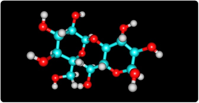
 Image Credit: Igor Petrushenko / Shutterstock.com
In eukaryotic cells, most proteins are modified after translation in ways such as the addition of glycans. These modifications can be essential for cell viability and function, such as cell differentiation, gene and metabolic regulation, protein activity, as well as pathogen recognition, cancer, and autoimmune reactions. Because of this, there is now an increased interest in using glycans as targets for diagnostics, therapeutics, and vaccines.
What techniques are best to study glycans?
Sugars molecules are not charged and do not absorb UV light or emit fluorescence.
Techniques such as High Performance Anion Exchange Chromatography with Pulsed Amperometric Detection (HPAEC-PAD) can be used to analyze glycans, or if they are modified to be fluorescent or methyl groups then liquid chromatography-mass spectrometry (LC/MS) can be utilized. If the glycan is abundant, then thin layer chromatography can be used, although the resolution from this method is poor.
In the study by Wheeler and co., which will be explored later, the authors used capillary electrophoresis and MALDI-ToF mass spectrometry to determine the sugars which were contained within glycosylated proteins.
What kind of modifications with glycans occurs?
Glycosylation can occur as a result of five types of glycosidic bonds; N-glycosyl, O-glycosyl, C-glycosyl, P-glycosyl, and glypiation. The N- and O-glycosyl bonds are the most common.
The N-glycosyl bonds are formed by GlcNAc sugar to the amino acid asparagine, and this is the most common protein-carbohydrate bond. This was initially described in the protein ovalbumin, and thereafter this particular bond has been recognized in other proteins, including plasma proteins, hormones, enzymes, cell surface receptors, immunoglobulins, and lectins.
O- glycosylation occurs where the carbohydrate unit is bound to an amino acid which has a hydroxyl group, such as serine, threonine, tyrosine, hydroxyproline, and hydroxylysine. O-glycosylation clusters attached to serine or threonine has been thought of as a component of mucins.
However, this has been shown in other proteins such as fetuin, human gonadotropins, glycophorins, and even in proteins within the S-layer of the archaebacterium Aneurinibacillus thermoaerophilus.
Combinations of N- and O-glycosylated glycans have been found in proteins such as fetuin, glycophorin, IgG immunoglobulin, insulin receptor and thyroid cell surface glycoproteins and mucins, proteoglycans, and collagens, which are thought to be carriers of carbohydrates bound through O-glycosylation.
How do proteins become glycosylated?
These glycan modifications are carried out by specific enzymes, and so far, enzymes that form 16 or so glycosyl-protein links have been identified. Typically, this occurs by the formation of a bond between an activated sugar or an activated sugar-phosphate unit and the target amino acid. Initially, this involves only one sugar unit, but others can be added subsequently to create oligosaccharide glycans.
How does glycosylation relate to disease?
Recent studies have shown that problems occurring during glycosylation can lead to disease. Sometimes, these can be congenital and mostly affect neurological development. The rare disorder leukocyte adhesion deficiency II has also been linked to a defect in protein glycosylation; in this case, the lack of GDP-Fuc formation.
Paroxysmal nocturnal hemoglobinuria is another disease associated with a defect in glycosylation, in this case, the addition of GlcNAc to the inositol residue of phosphatidylinositol.
Glycosylation and bacterial virulence
Bacteria of the genus Clostridium produce toxins and these act on mammalian Rho GTPases. The Rho proteins are molecular switches that control cellular processes, such as the organization of the actin filaments of the cytoskeleton. When GTP is bound, the Rho proteins are active, but the conversion of the GTP to GDP makes the Rho proteins inactive.
Modification by the addition of a GlcNac or Glc to a threonine residue by Clostridium toxins inhibits the activity of the Rho GTPases, which means that the proteins remain active. This, in turn, causes pathological states such as botulism, gas gangrene, and pseudomembranous colitis.
A recent study by Wheeler and co. showed that when the bacterium Pseudomonas aeruginosa is exposed to mucous, which contains mucin glycans, the expression of virulence genes is down-regulated, and that its biofilm becomes broken down.
Further, the authors found that exposure to mucins also promoted “planktonic” growth as opposed to biofilm growth. This perhaps is key to explaining how the mucous layer acts as the first barrier against infections.
Sources
neb.com What is Glycobiology? www.neb.com/applications/glycobiology-and-proteomics/glycobiology
neb.com FAQ: What are the best techniques to analyze N- and O- glycans? www.neb.com/.../what-are-the-best-techniques-to-analyze-n-and-o-glycans
Spiro, R. G. (2002) Protein glycosylation: nature, distribution, enzymatic formation, and disease implications of glycopeptide bonds. Glycobiology https://academic.oup.com/glycob/article/12/4/43R/590954
Wheeler, K. M and co. (2019) Mucin glycans attenuate the virulence of Pseudomonas aeruginosa in infection. Nature Microbiology https://www.nature.com/articles/s41564-019-0581-8
Further Reading
Last Updated: Nov 19, 2019