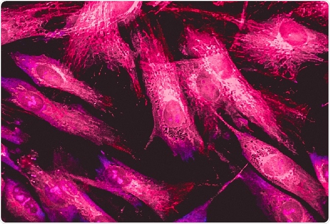
Wide-field fluorescence microscopy is a widely applied imaging technique used to examine cells and investigate their internal structures, including proteins and other cellular components.
 Image Credit: Vshivkova / Shutterstock
In this technique, the entire sample is illuminated and a 2D image of multiple proteins can be identified at once.
What are fluorophores?
Fluorophores are excited upon the absorption of a specific wavelength of light, resulting in the movement of electrons to a higher energy level. These electrons then fall to their original state, leading to the production of a longer wavelength. The absorbed wavelength is called the excitation wavelength, whereas the emitted wavelength is called the emission wavelength.
The Stokes shift defines the difference between these values. Each fluorophore can emit and absorb different wavelengths, which are known as their excitation and emission spectra. These ranges can used to define different types of fluorophore.
Sample preparation
Some biological samples naturally contain fluorophores (e.g. chlorophyll). Alternatively, biological samples can be labeled using different methods, including immunofluorescence, DNA manipulation, or fluorescent stains. Immunofluorescence uses fluorophore-connected antibodies, which recognize a specific antigen and therefore label the protein of interest.
DNA manipulation, however, genetically modifies the protein of interest to make it fluorescent. Stains can also be added to a sample which binds specifically to the protein of interest. For example, DAPI is a nucleic acid stain which identifies nuclei by binding to DNA.
Methodology
A lamp emits excitation light which passes through an optical filter which ensures that only specific excitation wavelengths fall on the sample. The light is then aimed at the sample via reflection off a dichroic mirror.
The excited fluorophores within the sample emit a higher emission wavelength that is detected by a camera. This data is then quantified and used to identify the specific location of the proteins within the sample.
The light source
Mercury or xenon arc-lamps are the traditional light sources used for this technique; however, they both produce very intense wavelengths which lead to bleaching and toxicity. Light-emitting diodes (LEDs) are typically used as they are cheap and compact, don’t lead to the bleaching and toxic effects can be accurately programmed.
High-resolution imaging
The resolution of a microscope determines how well a microscope can differentiate between different two closely placed objects. Although wide-field fluorescence microscopy has high resolution, imaging thick 3D samples remains difficult.
The entire sample is examined at once, and it can be challenging to identify the precise source of fluorescence. Background fluorescence can also increase the noise of the image.
In photobleaching, fluorophores react and covalently modify each other. This leads to a dimmer image as fluorophores are unable to emit fluorescence, reducing the resolution of the produced image.
Further Reading
Last Updated: Oct 24, 2018