What makes pre-clinical research so exciting?
To me the most exciting aspect of pre-clinical imaging is its broad range, from very basic science up to applied science. You deal with a range of disciplines including biology, chemistry, physics, biochemistry, biophysics, cell biology and of course medicine, as the aim is the translation of research to humans.
Every method or every therapy monitoring you develop in pre-clinical MRI can nearly always be translated to humans due to the non-invasive character of MRI.
Preclinical to Clinical
Preclinical to Clinical from AZoNetwork on Vimeo.
What projects are you and your team at the University of Freiburg currently working on?
Currently we have groups working with MRI in the main areas of neurology, oncology, cardiovascular and hyperpolarization.
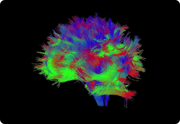
Our latest developments in the field of resting state fMRI (R-fMRI) and diffusion tensor imaging (DTI), combined with fiber tracking, are highly promising. Both techniques have applications in neurological questions.
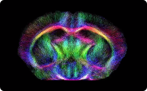
DTI allows you to measure the spatial resolution of the diffusivity of water especially within the brain. With this, you are able to calculate where your nervous fibers run, so you're able to get a map of the architecture of the brain.
What’s the main advantage of MRI?
The big advantage of MRI is its diversity in contrasts. You can basically tailor your sequence towards the task you have.
Whether you want to measure diffusion or perfusion; whether you want to know where does the brain work when you have a certain task; or whether you want to know how does the blood flow in the arteries.
What information does functional MRI provide you with in neurological studies?
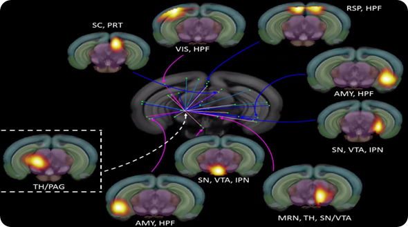
Functional MRI gives you information on the function of the brain. There, you have to differentiate between fMRI the classical functional MRI, and resting state MRI. Functional MRI shows you the active brain areas according to a certain stimulus.
In resting state fMRI, you don't have a stimulus, you record the activities of the brain over time and then derive from that information which areas within the brain communicate with each other.
Knowledge about structural and functional networks and their interactions allow us to study disease-related changes within the brain. Therefore, we yield a better understanding and thus treatment options for diseases like depression, addiction, or multiple sclerosis.
Please can you give some examples?
In addiction it is known, for example, that at the time and point you get addicted, there is a functional change within the brain, and a functional change always leads to a structural change as well. We currently search for those changes in an alcohol addiction model.
In multiple sclerosis, to give another example, loss of myelin occurs at the site of the lesion. We were able to detect this loss of myelin with our diffusion tensor imaging technique. We're currently investigating a treatment with an artificial hormone which should induce remyelination.
Talking about depression, a definition of affected areas could allow for targeted stimulation of those areas and this could drastically improve the life of people affected by this disease.
What are the latest technological developments that allow you to get such brilliant results?
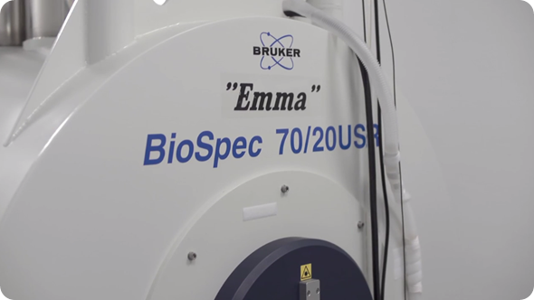
The most outstanding technological development helping us in the last years was the development of a cryogenically-cooled RF coil. This reduces the electronic noise drastically and thus leads to a gain in signal-to-noise ratio between a factor of two and three.
Only that higher sensitivity allows us to acquire high quality data in shorter time and only that yields the state-of-the-art Resting state and DTI data.
What are the clinical benefits of DTI and fMRI?
In the clinical world, knowledge about networks can certainly help in neurosurgery. Knowing which areas are critical can help neurosurgeons spare those areas during surgery. In addition, it might speed up the surgery.
Concerning our multiple sclerosis study, the pathway towards the clinic is clear. If we have positive results in our study, a clinical trial should follow directly.
What does the future hold for pre-clinical MRI? What innovations would you like to see in the field?
I believe that pre-clinical MRI has a bright future ahead. Not only because of the reasons we already discussed as investigating diseases, finding treatments, monitoring the treatments, and then translation, but also in the field of personalized medicine.
Take for example oncology, every tumor is different and every patient is different, and then the interactions between the both differ from patient to patient as well.
If you are able to take a specimen of the tumor of the patient, and test various treatments on it, you're able to tailor a treatment for that individual which will drastically improve the success rate of the treatment.
Where can readers find more information?
To find out more, visit the Molecular Imaging Research Center website.
About Prof Dominik von Elverfeldt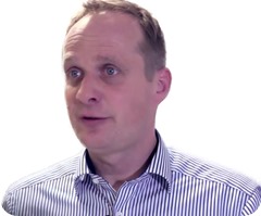
Prof Elverfeldt is currently group leader of the Advanced Molecular Imaging Research Center at the University Clinic Freiburg.
Originally a physicist, Prof. Elverfeldt has since moved into the pre-clinical MRI field, which covers a broad range of disciplines.