After the first ever MediSens conference, 2016, it is fair to say that there is much excitement in the area of medical imaging. The industry is experiencing a drive towards a more personalized approach to healthcare, providing patients with treatment that is specific and adaptive to them.
The conference was sponsored by some of the leading names in molecular imaging, including ON Semiconductor, Hamamatsu, Siemens Healthcare and PerkinElmer, all keen to show off their latest developments in imaging technology.
For example, ON Semiconductor were promoting their image sensors from medical applications from exceptionally small CMOS image sensors for use in endoscopes, to high quality performance CCDs for use in x-rays.
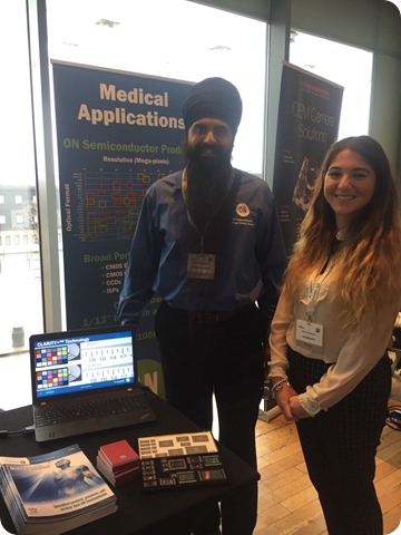
With Roopinder Grewal, Senior Director, ON Semiconductor
The need for multimodality imaging in healthcare was first introduced at the event by Dr Dimitra Darambara, from The Institute of Cancer Research, who compared imaging in cancer to the Hanabusa Itchō artwork titled ‘Blink monks examining an elephant’.
Dr Dimitra explained the story of how each monk felt a different part of the elephant and therefore described the elephant’s appearance differently to one another. To apply this analogy to imaging – we have many different ways to look at a disease such as cancer, but each modality gives us a different perspective.
These pieces are correct individually, but they do not provide the whole picture, so going forward there is a need to join together multiple modalities in order to quantitatively look at cancer in patients.
Dr Darambara summarized with Hippocrates’ quotation:
It is more important to know what person the disease has than what disease the person has”
This provoked thoughts of how we can increase the quality of medical imaging in order to allow for interpatient heterogeneity.
This was further expanded during Dr Antonis Kalemis’ talk. The Vice President of the Association of Imaging Producers & Equipment suppliers, explained that in the modern world, we are seeing a move towards hybrid technology systems in order to image cancer quantitatively.
This way, it is possible to measure exactly how much radiology activity you have in a patient. As an example, Kalemis gave us 4 systems that show the future direction of SPECT, with technology from Molecular Dynamics (Valiance X12), MILabs (G-SPECT), GE Healthcare (Discovery NM/CT 670 CZT) and Siemens (Symbia Intevo xSPECT).
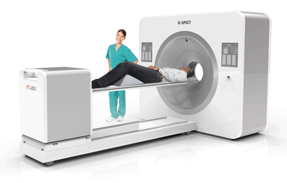
G-SPECT, from MILabs
The hybrid systems mean that not only is it the tumor that can be identified, but there is also the ability to trace the tumor and see if/when it becomes less metabolically active. This has potential applications towards adaptive patient healthcare, as it means that HCP’s can use this to make a decision on: are the side effects on the patient worth the impact the treatment is having on the tumor? If not, then it may be more appropriate to try a new method, and perhaps this treatment would be better suited for a patient with a different genetic makeup.
Discovery NM/CT 670 CZT introduction video, GE Healthcare
Dr Kalemis also presented a number of challenges molecular imaging faces when it comes to medical applications, some of which at first thought, are not obvious. Macro-environmental factors, for example demographics, are putting pressure on technology advances in healthcare.
In this example, an increase in population, life expectancy and quality of life mean that there are more patients, illnesses are prolonged over longer periods of time and there are raised expectations for treatments, respectively. This suggests that industry leaders in developing new technology for medical imaging are under tremendous pressure, and are doing an exceptional job to keep up with this.
Dr Phil Evans, Professor of Medical Radiation Imaging, University of Surrey, moved the topic of conversation to proton therapy. Protons travel straight through the patient and come out of the other side.
The proton can then provide information of its route along the way, hence allowing you to look at the effect the patient’s tissue has on the proton. The PRaVDA (Proton Radiotherapy Verification and Dosimetry Applications) integrated imaging system has been developed for this purpose, and could be very valuable to HCP’s in assessing how the patient is responding to the treatment.
A different technology ready for x-ray medical imaging called CMOS (complementary metal-oxide-semiconductor) was introduced by Dr Biju Jacob, Senior Engineer in X-ray Detector development at General Electric. Dr Jacob highlighted that the OEC Elite CFDTM utilises CMOS flat panel detector technology resulting in better image quality even at low dose, and has a high dynamic range.
Dr Jacob’s team at GE confirm that:
CMOS technology pushes the limits of x-ray detection with improved sensitivity, enhanced spatial resolution and speed. CMOS based flat panel detectors offer an excellent alternative to image intensifiers in the mobile C-arm systems”
The idea of CMOS touches on how we are able to explore alternative technologies in order to improve healthcare through better imaging.
Dr Sandro Francesco Tedde, Key Expert Research Scientist, presented some interesting findings from research at Siemens Healthcare with hybrid x-ray detectors, which really demonstrates passion towards testing the suitability of a new technology to medical imaging.
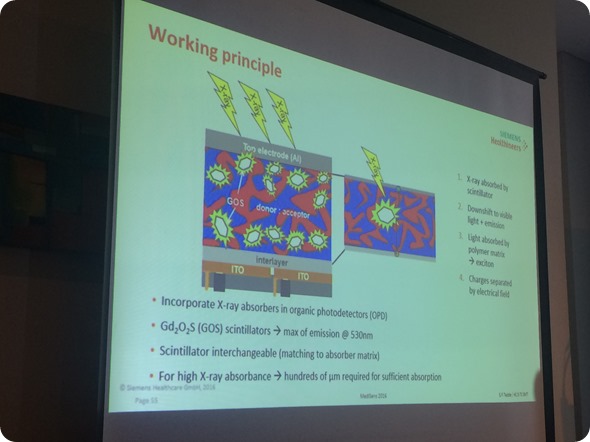
It was found that these hybrid x-ray systems have a better lens performance than conventional systems, described as Modulation Transfer Function (MTF), resulting in a higher quality image. Although the system still faces issues, for example thick devices showing a lack in sensitivity, this is something that can be worked on, and therefore the hybrid organic photodetectors have a huge potential to become the next generation technology for flat panel x-ray detectors.
Professor Gary Royle, Professor of Medical Radiation Physics, UCL, gave an engaging talk on what we need to image during treatment. What it is easy to forget, is that as the patient is living and breathing in real time, the tumor is moving with them, especially if placed near the diaphragm.
It would be counter-productive if during radiotherapy, the treatment hits and causes damage to surrounding tissue. Therefore, it is essential to know how the patient moves in order to track and hit the tumor as it moves.
Prof Royle also touched on how it is important to change the treatment as the patient changes, for example as the patient loses weight due to the treatment, their movements and therefore their tumour movements will change. This reiterates the need for future adaptive radiotherapy, and the importance of accurate medical imaging in order to accurately perform treatment procedures.
MediSens Conference 2016 - Highlights and testimonials
Despite the pressures on technology in medical imaging, it is extremely inspiring as well as comforting to see so much effort and research going into making healthcare more adaptive for patients through imaging technologies.
 Scientists turn red blood cells into long-lasting drug and imaging carriers in mice