An interview with Dr Susan Tappan, explaining MBF's partnership with Huron Digital Pathology allows researchers to achieve large-scale, repeatable whole slide imaging, conducted at SfN 2018 by Alina Shrourou, BSc.
What is meant by “whole-slide” imaging?
Whole slide imaging is the capability to scan at high resolution an entire glass slide that has multiple histological sections cut with a microtome, either imaged with brightfield or florescence. Whole slide imaging can be used to advance many aspects of biological research because it can serve as a faithful archive of sensitive or precious archival material, either human or high-quality experimental material. You can create an image representation that can be used for analysis, exploration, and communication because it's able to be sent and viewed across the internet, which expands the reach of these very precious glass slides.
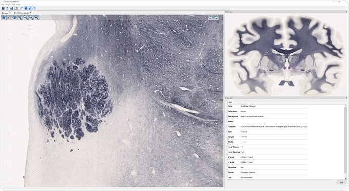
Image credit: MBF Biosciences
How can whole-slide imaging be used to advance biological research? How does it compare to traditional glass slides?
In day-to-day research where you have hundreds of glass slides that may make up a typical experiment, being able to scan the slides into whole slide image allows you to catalog , analyse, and return to them at any time for additional analyses as new insights are gained from other aspects of your research. Therefore, it provides a mechanism for you to get more out of your data in a very facile way.
How have MBF recently partnered with Huron Digital Pathology? And how will this help life sciences researchers achieve large-scale, repeatable whole slide imaging workflows?
We're very excited to partner recently partnered with Huron Digital Pathology. Huron leads the way in solutions for pathology research. Huron Digital Pathology make a very high-quality image scanner. The scanner itself is very fast, and produces beautiful alignment and images.
It's also very easy to use. It fits very nicely into our product offerings which allow researchers to extract quantitative data from their whole slide images, either from doing stereology, which is an unbiased mechanism for quantifying biological features, or doing medical education and 3D reconstruction of features of interest. Huron has also been very receptive to our input, based on decades of experience in performing quantitative analysis for research. This partnership allows us to deliver the very best state-of-the-art technology for research.
Please give an overview of MBF’s software and how this interacts with Huron’s TissueScope.
By partnering with Huron we've provided a streamlined process to go from a glass slide to a whole slide image, and then to store that in the cloud using our Biolucida image server.
That server can be based at your institution, or hosted on AWS or similar cloud service provider. The images can be utilized in our analytical software, Neurolucida 360 or Stereo Investigator, to get repeatable, reliable quantitative measures from your images.
Alternatively, researchers can utilize our NeuroInfo software in order to identify individual neuroanatomical regions by automatic registration with the Allen Institute Mouse Brain Atlas or reconstruct 3D image volumes from serial section images using our TissueMaker or BrainMaker software.
Neurolucida
How do you think this collaboration will advance pathology research?
I think that the ability to scan glass slides to create a whole slide image at a high enough resolution to get cellular or subcellular detail is highly relevant for pathology applications. The ability to share the image data with your colleagues at any point really expands the ability of the glass slide to leave your laboratory and actually be approachable by other researchers. Also, our expanding capabilities to provide automated, artificial intelligence- driven, big-data analyses of large images makes the use of Huron scanners even more important.
What are MBF bringing to the scientific community at Neuroscience 2018?
MBF is hard at work to bring reliable, sophisticated quantitative analyses to the emerging imaging technologies, that really push the envelope for neuroscience research. This includes everything from expansion microscopy, to STED, to whole slide imaging. We're very interested in image analysis applications across the breadth of the neuroscience communities.

About Dr. Susan Tappan
Susan Tappan, Ph.D. is the Scientific Director at MBF Bioscience. She has a broad interest in neuroscience with a focus in neuroanatomy.