To gain further insight into the brain and its functions, it is necessary to atlas the brain and the connections involved. However, this large scale task is a difficult and time-consuming process. The demand for high quality images places greater strains on image acquisition and management, with factors such as cross-modal compatibility, visualization and imaging speed requiring consideration. As a part of advancing neuroscience research, the US and EU depend on developments in IT and imaging to make this task more feasible.
Whole-mount digital slide imaging plays an important role by providing high resolution for detailed, integrated models of the brain. Making the generation of whole brain models and simulations a reality depends on the ability to produce high quality images rapidly and combine them with other modalities. Although the creation of reference brains is on the increase, the use of new imaging technology is required to expedite this process. Huron Digital Pathology’s TissueScope™ solution provides several benefits to researchers creating human brain models, thereby enabling this process to be faster and more feasible than ever before.
BigBrain Project
The BigBrain project at the Jülich Research Centre in Germany has led to immense innovation with respect to the generation of reference brains. Led by Katrin Amunts, BigBrain is a free-access 3D reference brain that used whole-mount slide scanning to provide detailed mapping, at an almost cellular level. Before this project, only macroscopic levels of detail, with 1mm resolution, had been achieved for reference brains.
The innovative BigBrain project enables researchers to visualize the borders between higher associative areas and primary cortical areas. Previously, traditional two-dimensional slide scanning had been used to achieve this, but this was restricted to only sections of the brain that researchers chose to slice. However, using whole-mount slide scanning to create a 3D reference brain, the whole border of the cortical ribbon can be defined. Likewise, 3D analysis of the individual variations between macroscopic landmarks and cytoarchitectonic borders, has not been possible before. The ability to map functional probability maps to the BigBrain data set means cross-modal research is possible. As a result, brain activity maps, neural projections, and gene expression maps can all be related to cytoarchitecture. All of these new capabilities mean researchers can now validate new hypotheses on the distribution of, for example, fiber bundles, transmitter receptors and genetics.
The procedure through which Amunt created BigBrain shows the logistics involved in digital slide scanning, as well as the potential scope for improvement. The process involved embedding the specimen in paraffin and splitting it into 7404 slices that were 20μm thick. The slices were then scanned for over 1000h at 10μm resolution and then downscaled to achieve an overall isotropic resolution of 20μm. The dataset size was one Terabyte in total. Although, models with an even higher resolution are in demand, the projected size of such high resolution data does present computing challenges in terms of being able to actively explore this amount of data rather than simply store it. Moreover, additional data sets that are similar to BigBrain need to be produced because, despite being very powerful, BigBrain only represents the brain of a 65-year old female and researchers need to further analyze the individual variability in the architecture of cortical borders.
Huron’s TissueScope™ Solution
Given that additional reference brain data sets of similar or even better image quality are required, researchers are asking how this procedure can be improved and whether a balance can be struck between the scanning time and image quality. Some investigators are wondering whether thicker optical z-stacks, enabled via chemical techniques, could be a better technique. These problems and more have been addressed by Huron Digital Pathology’s TissueScope™, with its high quality and high efficiency scanning making it well suited for the creation of reference brains.
The key benefit of TissueScope™ is its ability to scan both small and large specimens, with sizes ranging from 1 x 3” up to 6 x 8” (Figure 1). As a result, the scanning time is instantly reduced and the need for stitching and tilting is eliminated. The user interface allows scanning of the specimen at different magnifications including 1x, 2x, 10x, 20x and 0.25μm at 40x, therefore enabling easy imaging of the areas of interest.
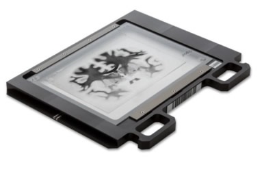
Figure 1. A brain specimen on a 5 x 7” slide in Huron’s whole-mount slideholder. Image credit: Huron Digital Pathology.
In addition, TissueScope™ can be used to acquire z-stacks and optical sections on specific ranges of tissue up to 500μm or even 2mm in depth using the appropriate clearing methods. Use of the optional TissueSnap™ workflow accessory avoids the need for pre-processing and preview scanning, meaning the main scanner can operate at full throughput and the time to view is further reduced. The scans performed using a Huron scanner are stored in non-proprietary, open .tiff format, which enables researchers to use a range of image analysis software to create 3D reconstructions of tissue (Figure 2).
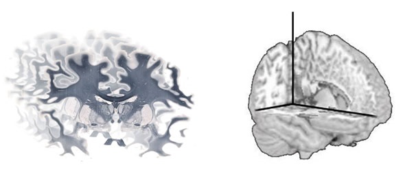
Picture 2. A series of whole brain images can be reconstructed using software to produce a 3D volume. Image credit: Huron Digital Pathology.
The time BigBrain took to acquire data for all 7404 images was 1000h. Overall, it took five years to develop BigBrain, with one year dedicated to data acquisition and the other four years to constructing the 3D volume. By contrast, the TissueScope is able to scan a whole brain slide at 1μm resolution, in just 12min, suggesting that future reference brains using TissueScope could achieve a resolution 400x higher than the current BigBrain and acquire these images within six months.
Huron Digital Pathology has already supported various human brain research projects by enabling high resolution, whole brain images to be acquired.
About Huron Digital Pathology
 Based in Waterloo, Ontario, Canada, Huron Digital Pathology has a 20 year history designing sophisticated imaging instrumentation. Our end-to-end digital whole slide imaging solutions for digital pathology incorporate our award-winning TissueScope™ digital slide scanners; TissueView™ image viewing, sharing and management platform; and our workflow-enhancing accessories, which include our innovative TissueSnap™ preview scanning station.
Sponsored Content Policy: News-Medical.net publishes articles and related content that may be derived from sources where we have existing commercial relationships, provided such content adds value to the core editorial ethos of News-Medical.Net which is to educate and inform site visitors interested in medical research, science, medical devices and treatments.