The quick encapsulation of individual cells in miniature, picoliter-volume, water-in-oil droplets (picodroplets) is the foundation of the revolutionary single-cell analysis method known as picodroplet technology.
This strategy makes it possible to conduct research at the single-cell level much more effectively than with conventional approaches1 by enabling high-throughput screening, gentle cell processing, and high-sensitivity quantitative tests.
The capacity to screen individual cells in their natural microenvironment with better accuracy and sensitivity is made possible by the use of picodroplets, which function as small test tubes.
With the help of this format, cells can be protected from shear stress during subsequent analysis and preserved in a unique protective environment to promote their viability and integrity during incubation.
Leading pharmaceutical organizations are already utilizing this strategy to improve the workflows for antibody identification and cell line development and to hasten the discovery of new biotherapeutics after realizing the promise of picodroplet technology.
However, a broader range of other fields and applications, including cell therapy and genome editing, can benefit from picodroplet technology.
Dilution is reduced when a cell is contained within a picodroplet, and chemicals generated by the cell will accumulate and reach a measurable concentration in a comparatively short amount of time.
This often happens in applications like antibody discovery after 1–4 hours of incubation. Such downsizing might enhance the transport of reagents and aid in the identification of molecules produced by individual cells after a successful editing event in genome-editing applications.
Picodroplet compartmentalization guarantees that genetic editing occurs on a single cell as opposed to a large population of cells. Additionally, compartmentalization allows for easier detection of low-frequency events, supporting applications such as the sorting of rare genome-edited cells.
Therefore, picodroplet applications for genome editing can offer a number of benefits. For it to work, however, long-term cell incubation in picodroplets is necessary.
This article illustrates the viability and adaptability of picodroplet technology for uses requiring extended incubation durations.
Aims and objectives
The study outlined in this article demonstrates that:
- Different cell lines can survive in picodroplets for various incubation periods
- Suspension cells thrive in picodroplets
- After regrowth from picodroplets, single cells can form colonies
Method
A series of experiments were carried out to evaluate if picodroplets support cell viability, proliferation, and consequential clonal outgrowth of single cells following a 24-72 hour period of encapsulation in picodroplets.
In parallel testing, seven distinct cell lines - HCT116, MCF7, A431, A549, and HEK293 for adherent lines and Raji, THP1, Jurkat, and CHO-K1 for suspension lines - were used as models for both suspension and adherent cells.
Single cell encapsulation
The input cell concentration (2e5 cell/ml) was selected to maximize the number of picodroplets that would subsequently be generated, consisting of a single cell.
Using PicoSurf™ 1 surfactant and a Pico-Gen™ 1 Biochip, cells were suspended in culture media and enclosed in 450 pL picodroplets. The emulsion was then collected in a 1.5 mL Eppendorf tube and incubated in a CO2 incubator.
Four independent time points - after cell encapsulation (time 0) and after 24, 48, and 72 hours - were used to examine the vitality and proliferation of the cells.
Cell viability
Acridine Orange and DAPI staining were used to determine the viability of the cells (Acridine Orange stains all live and dead cells, allowing for a complete cell count, whereas DAPI will only stain the dead cells).
Pico-Break™, a reagent that lowers picodroplet stability, was used to break the picodroplets at each time point after a sample of the emulsion was obtained. After then, a portion of this cell suspension was dyed with Acridine Orange and DAPI and examined with a Nucleocounter NC-3000 (Figure 1).
Based on this study, a portion of the material that had not been stained was planted in 96-well plates to be examined for outgrowth.
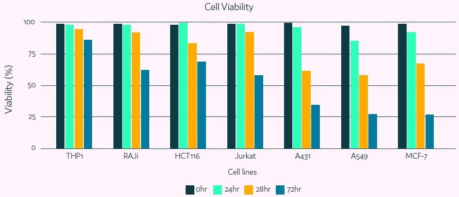
Figure 1. Cell viability in picodroplets. Image Credit: Fluidic Sciences and Sphere Bio
As shown in Figure 1, after 24 hours, all but one line (A549) still showed viability over 90%. At 48 hours, more lines’ viability began to fall, with adherent lines A549, A431, and MCF7 suffering the most.
The pattern is still present after 72 hours of picodroplet incubation, with viability from the A549, A431, and MCF7 cells dropping below 40%; Raji, HCT116, and Jurkat cells exhibit vitality around 60%; while THP1, CHO-K1, and HEK 293 cells show the viability of 80% and higher.
These studies showed that cell viability is influenced differentially depending on the cell line after picodroplet encapsulation and up to 72 hours of incubation, with suspension lines often outliving adherent ones.
However, the majority of cell lines showed a respectable capacity for survival in picodroplets for 24–48 hours, suggesting the potential viability for applications requiring extended incubation. It can be said that the lack of nutrients is mostly to blame for the observed decline in cell viability.
Cell proliferation
To establish if proliferation took place within the picodroplets, the concentration of surviving cells was compared to the initial concentration using the count from the cell viability experiment.
The outcomes showed a rise in cell concentration, notably for the suspension cells Raji, THP1, Jurkat, and CHO-K1, proving that picodroplets can promote specific cell lines’ potential for proliferation (Figure 2).
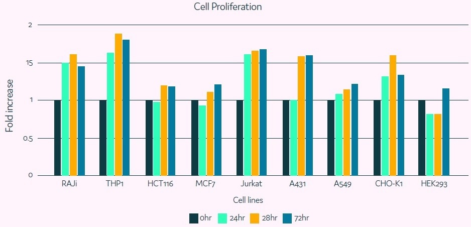
Figure 2. Cell proliferation in picodroplets. Image Credit: Fluidic Sciences and Sphere Bio
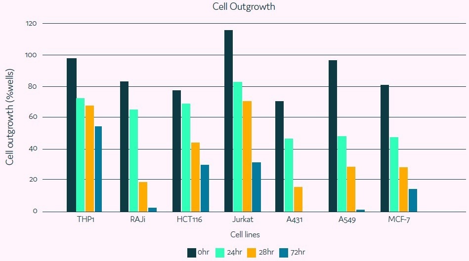
Figure 3. Cell outgrowth after incubation in picodroplets. Image Credit: Fluidic Sciences and Sphere Bio
This possibility could serve as a basis for further method development that will enable the generation of a “clonal population” already inside the picodroplet.
 Download the full paper
About Fluidic Sciences and Sphere Bio
Fluidic Sciences develops transformative in‑solution technologies for protein interaction analysis. Its flagship Fluidity One‑M instrument leverages Microfluidic Diffusional Sizing (MDS) to measure binding affinity, stoichiometry, size, and concentration without immobilization - directly in complex backgrounds such as serum, plasma, and lysate.
Sphere Bio is a brand of Fluidic Sciences. Its technology develops and manufactures single‑cell analysis and monoclonality assurance systems that enable researchers to find, analyze, and isolate the most valuable cells with speed and precision. Its proprietary picodroplet microfluidics and Cyto‑Mine® Chroma multiplexing platform power applications across antibody discovery, cell line development, cell engineering, and cell therapy.
Sponsored Content Policy: News-Medical.net publishes articles and related content that may be derived from sources where we have existing commercial relationships, provided such content adds value to the core editorial ethos of News-Medical.Net which is to educate and inform site visitors interested in medical research, science, medical devices and treatments.