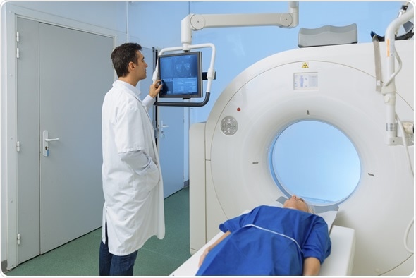
Magnetic resonance imaging (MRI) is an imaging technique which physicians use for the diagnosis and treatment of medical conditions.
The main components of this procedure are a magnetic field, radiofrequency pulses, and a means by which the detailed images can be studied.
Ordinarily, a specialized computer is used to view the images of internal anatomical structures.

MRI Machine - Image Copyright: s4svisuals / Shutterstock
Process
An MRI scan utilizes magnets, which are responsible for producing and exerting a strong magnetic field. When a patient is undergoing the scan, the hydrogen atoms (protons) in their body become aligned with the magnetic field, i.e., the net magnetic moment of the protons is parallel to the direction of the field.
When the patient is then subjected to a radiofrequency pulse, which is directed perpendicular to the magnetic field the protons undergo a change in magnetic moment, tilting away from the direction parallel to the magnetic field.
Once this radiofrequency pulse is removed, the protons’ net magnetic moment realigns with the magnetic field. This process involves a return to equilibrium for the protons – it is called relaxation.
How does MRI work? Jerome Maller explains
The energy release during this results in signals, which eventually lead to the construction of MR images. The time relaxation in addition to the quantity of energy released is reliant on the chemical milieu and intrinsic characteristics of the molecules.
Radiologists will differentiate between the tissue types based on the tissue-specific magnetic properties.
Administration of contrast agents (the majority of which contain Gadolinium) intravenously before or during the MRI can speed up the process of protons realignment. This results in brighter MRI images.
Procedure
The patient must lie on an examination table which is then inserted into the MRI unit. The examination is performed by a radiologist and technologist via a computer located outside the MRI room with the patient.
Any required contrast agent for the imaging session will be intravenously injected into the patient’s arm following an initial set of scans.
The MRI exam will generally include taking cross-sectional image sequences which may last as long as several minutes, with the entire examination possibly taking 30 - 50 minutes.
Limitations
- Patients must be able to remain still – this involves holding their breath during the procedure in order for high-quality images to be obtained. Naturally, this may be difficult to attain if the patient is nervous or suffering from pain.
- Not all MRI machines will be appropriate for a patient – a patient may be too large to fit in some forms of the machines.
- Good quality images may not be achieved if the patient has metallic medically necessary objects, e.g. an implant.
- Breathing may cause artefacts or image distortions during MRIs of areas such as the chest, pelvis and abdomen. Similarly, bowel motion can cause artefacts in abdomen and pelvic MRI images. Fortunately, the introduction of state-of-the art scanners and improved techniques allows these obstacles to be better overcome.
- Despite no evidence indicative of fetal risk, pregnant patients are not encouraged to undergo an MRI examination within their first trimester unless it is deemed as necessary.
- MRI might not prove effective in distinguishing between cancerous and edematous tissue.
- MRI is not as cost-effective as other imaging procedures and it may also consume more time.
References
Further Reading
Last Updated: May 18, 2023