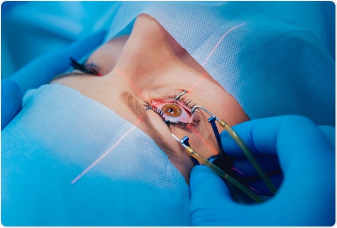
Laser in situ keratomileusis (LASIK) was initially reported by Ioannis Pallikaris in 1990 and has since become an efficient and highly popular procedure in corneal refractive surgery. However, a reduction of visual performance and the occurrence of higher-order aberrations, such as glare and halo under dim conditions, decrease of contrast sensitivity, and poor night vision, have been reported after the surgery.
The introduction of wavefront-guided laser technology in 1999 was a significant step forward for the field of refractive surgery, as it allowed for the correction of both spherocylindrical errors and higher-order aberrations. Hence, this surgical procedure was a great candidate for reducing post-LASIK night vision problems and improved resulting vision acuity.

Image Credit: Roman Zaiets / Shutterstock.com
Fundamentals of the technology
Wavefront-guided LASIK represents a refinement of classical refraction methods where the laser correction is tailored to the specific pattern of corneal refractive aberration in each patient. As the cylindrical aberration in corneas is not always uniform, applying a homogeneous correction over the entire area may not give the optimal result.
The surgeon begins the procedure by using the wavefront device to transmit a safe ray of light into the eye. The light gets reflected back off the posterior portion of the eye and goes out through the pupil before it finally reaches the aberrometry device, where the spherocylinder refraction is calculated based on a 4 millimeter (mm) entrance pupil. The reflected wave of light is then arranged into a unique pattern that measures the sphere and cylinder of the eye, as well as the higher-order aberrations.
Customized wavefront-guided Treatments (LASIK & PRK) (English)
Acquired visual measurements are then displayed as a three-dimensional (3D) map, also known as a wavefront map. Nomograms to independently adjust the amount of sphere and cylinder treatment should also be developed. The formatted and processed information can then be electronically transferred to the laser and computer-matched to the position of the eye, allowing the specialist to personalize the LASIK procedure for specific visual requirements of each patient.
The VISX WaveScan system was the first laser-based technology to be approved by the United States Food and Drug Administration (FDA) for the treatment of myopia (nearsightedness) and astigmatism. This system, which measures the refractive error and wavefront aberrations of the human eye using a Shack-Hartmann wavefront principle, consists of the S4 excimer laser and the WaveScan wavefront device.
Potential challenges
Wavefront-guided laser ablations are superior to conventional LASIK surgery, with some authors indicating that the end result of this procedure can even be “supervision.” Ocular or total wavefront-guided ablations have demonstrated effectiveness in minimizing aberrations without previous unsuccessful or non-optimized refractive eye surgeries. Still, this technique is not appropriate for every eye.
There are some unique challenges and obstacles for the application of wavefront technology. The human eye is a dynamic system with constant biological fluctuations, such as changes in pupil size and focus; therefore, the eye significantly differs from a telescope, which is static in nature. Although wavefront represents an excellent mapping system, it does not necessarily mean that the result will be the exact replica. In other words, ablating corneal tissue is not nearly as precise.
Wavefront-guided ablations may result in the removal of more tissue as compared to conventional ablations, depending on the individual patient. In cases of thin corneas, for example, this could be an issue. In addition, the benefits of wavefront-guided treatment may be masked by epithelial hyperplasia in such surface ablations.
In conclusion, when considering any type of corneal-based refractive surgery (LASIK, Epi-Lasik, or photorefractive keratectomy), a wavefront diagnostic should be considered. This approach will help to determine if critical higher-order aberrations are below normal, normal, or elevated. If they are elevated, either wavefront-guided LASIK is required, or no surgery is suitable.
References
Further Reading
Last Updated: Nov 29, 2022