Cathepsins are enzymes (known as proteases) that break down proteins. They play an important role in cell turnover, which is the process by which cells are replaced by young cells when older cells die.
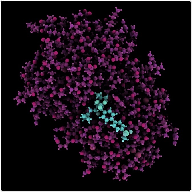
 Cathepsin K enzyme bound to the inhibitor odanacatib. Image Credit: StudioMolekuul/Shutterstock.com
They are mostly active within a cell’s organelle, but some operate outside of the cell. They are present in all animals, including humans, and have the potential to function as targets for therapeutic drugs.
Cathepsin inhibitors that block the activity of certain cathepsins have been the focus of studies that investigate treatment for a wide range of human diseases.
Cathepsins are implicated in several diseases, such as cancer, Alzheimer’s disease, arthritis, and some eye diseases, including age-related macular degeneration and glaucoma. Cathepsins have also been found to be active in viruses such as Ebola and severe acute respiratory syndrome (SARS).
The types of cathepsins
Cathepsins are organized according to their structure, which proteins they are responsible for breaking down, and by their catalytic mechanism.
Cathepsins are grouped as follows:
- Cysteine proteases B, C, F, H, K, L, O, S, V, W, and X – these cathepsins degrade proteins and are often found in fruits such as pineapple, fig, and papaya. In humans, they are involved in apoptosis (programmed cell death).
- Aspartyl proteases D and E – cathepsin D is unique in that it can be active at neutral pH, and it can activate certain growth factors. It has been implicated in tumor progression in cancer; cathepsin E is found in the skin and immune cells in humans and is believed to play a part in age-related neuronal cell death.
- Serine proteases A and G – these cathepsins break down peptide bonds in proteins, and low levels of cathepsin A can be associated with galactosialidosis, but its levels are increased in muscles that have been affected by dystrophy. Cathepsin G is involved in rheumatoid arthritis and infectious inflammatory diseases such as cystic fibrosis and acute respiratory distress syndrome (ARDS). Cathepsin G may also be a potential target for pain control.
Some cathepsins are found throughout the human body (cathepsin B, H, X, O, C, and L), but the rest are found in specific tissues or cell types. For instance, cathepsin F is found in macrophages, K is found in osteoclasts, S in antigen-presenting cells, V in thymus cells, and W is found in both natural killer (NT) cells and cytotoxic (CTL) cells.
The types of cathepsin inhibitors
Pepstatin is a peptide inhibitor, and pepstatin A is widely documented as being an effective inhibitor of cathepsin D and E, although its effect on cells as a drug is not yet understood.
Cathepsin A and G are inhibited by being paired with their serine protease inhibitor, which blocks their activity when it is not needed. Synthetic inhibitors that are often used in research into therapeutic drugs can also inhibit cathepsin A and G, and 4-benzenesulfonyl fluoride hydrochloride (AEBSF) and phenylmethylsulfonyl fluoride (PMSF) are also used to inhibit serine proteases.
Antithrombin and alpha 1-antitrypsin (A1AT) are serine protease inhibitors that have been widely investigated for their role in human diseases. One A1AT disorder is characterized by a hereditary deficiency in A1AT, which leads to the chronic breakdown of tissues that largely affects the lungs.
Although A1AT deficiencies typically affect the lungs, there has been evidence to suggest that the protein has anti-inflammatory roles outside of the lungs, as antibodies against A1AT have been found in the synovial fluid in people living with rheumatoid arthritis. These A1AT antibodies have been linked to more severe RA and are present long before the onset of the disease.
A1AT is also associated with liver cirrhosis, a condition in which the liver tissue is replaced with scar tissue due to long-term damage, because of a mutation in the genes responsible for encoding A1AT.
In cancer, lower levels of cathepsin inhibitors have been linked to more aggressive disease, as increasing the activity of certain cathepsins can facilitate the movement of malignant cells from their primary site to secondary sites.
There are several roles that specific cathepsins play in cancer progression. Cathepsin D has been found to reduce the immune system’s anti-tumor response, and cathepsins B and L are heavily involved in cell invasion. Using cathepsin inhibitors stops cell invasion and subsequent metastasis, thereby reducing the risk of cancer spread.
Natural cathepsin inhibitors
Several natural products are inhibitors of certain cathepsins, and natural products are often the first area of study for those looking into potential drug candidates for a wide range of diseases.
These cathepsin inhibitors can be extracted from plants, fungi, and bacteria. Such natural cathepsin inhibitors include:
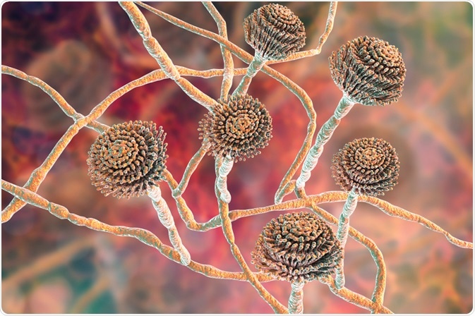
- Aspergillus – inhibits cathepsin B
- Marine cyanobacterial metabolite gallinamide A – inhibits cathepsin L
- Amino acid derivatives – inhibit cathepsin A
- Bifalvones – inhibit cathepsins B and K
- Chalcone products
 Fungi Aspergillus. Image Credit: Kateryna Kon/Shutterstock.com
Summary
Cathepsin inhibitors block the activity of a range of enzymes or cathepsins that are found throughout the human body. Certain cathepsins are associated with more aggressive disease and poorer prognosis in some types of cancers, and they have also been found to be present in the body before the onset of certain diseases such as rheumatoid arthritis, which may help in identifying the risk of disease in patients before symptoms occur.
Cathepsin inhibitors are proving to be a promising area of study regarding the development of therapeutic drugs for an array of common human diseases, and regulating the activity of cathepsins may be used as a target to treat or manage these diseases.
References
- Alitalo, K. et al., KLK3/PSA, and cathepsin D activate VEGF-C and VEGF-D. Cancer Biology. https://elifesciences.org/articles/44478
- Bradshaw, R. A., Stahl, P. D., eds., (2016). Encyclopedia of Cell Biology, Volume 1. Massachusetts, USA: Academic Press. p.681.
- Breugelmans, B., et al. Pacifastin-related peptides: Structural and functional characteristics of a family of serine peptidase inhibitors. Peptides.
- González, F. V., Vidal-Albalat, A. Natural products as cathepsin inhibitors. Studies in Natural Products Chemistry. https://doi.org/10.1016/B978-0-444-63749-9.00006-2
- https://www.sciencedirect.com/science/article/abs/pii/S0196978108003240?via%3Dihub
- Im, E., Kazlauskas, A. The role of cathepsins in ocular physiology and pathology. Experimental Eye Research. https://doi.org/10.1016/j.exer.2006.05.017
- Katunuma, N., Nomura, T. Involvement of cathepsins in the invasion, metastasis, and proliferation of cancer cells. The Journal of Medical Investigation. https://doi.org/10.2152/jmi.52.1
- MEROPS. (2017). https://www.ebi.ac.uk/merops/inhibitors/index.shtml
- Yamashima, T. Reconsider Alzheimer’s disease by the ‘calpain-cathepsin hypothesis’: a perspective review. Progress in Neurobiology. https://doi.org/10.1016/j.pneurobio.2013.02.004
Further Reading
Last Updated: Mar 4, 2021