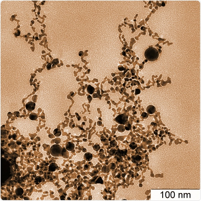
Gold nanoparticles are between 1–100 nm in diameter and show great potential as drug delivery vehicles, contrast agents, biosensors, and radiation dose enhancers. However, basic understanding of how the physical and chemical properties of these particles interact with a biological environment is lacking.
Upon intravenous administration, nanoparticles must overcome opsonisation and clearance by reticuloendothelial system, locate tumors, penetrate target cells to deliver drugs, and be safely eliminated from the body without bioaccumulation. The ability to bypass these obstacles is dependent on the physical dimensions and chemical properties of the nanoparticle.

Gold nanoparticles of various shapes obtained by laser ablation in water. Image Credit: Georgy Shafeev / Shutterstock
Nanoparticle Surface Chemistry
Proteins present in the blood accumulate on foreign bodies, such as nanoparticles for uptake and excretion by phagocytotic cells. The formation of these proteins around the nanoparticle is known as a protein corona prevents the drug delivery functions of nanoparticles.
Poly-ethylene glycol (PEG) is commonly used to coat nanoparticles for in vivo use, as it is biocompatible and prevents the formation of the protein corona. Overexpressed membrane proteins on cancer cells can be exploited by targeting complimentary biomolecules which can be internalised along with the nanoparticle by active transport mechanisms.
Very large nanoparticles, or those that have become several times larger than they were initially due to the formation of the protein corona, have difficulty passing through the renal filtration system of the liver.
Alternatively, these particles may be excreted by the hepatobiliary system, which takes significantly longer. Zwitterionic coatings encourage renal clearance more effectively than positively or negatively charged coatings, while also offering better resistance to protein accumulation.
Gold Nanoparticle - Sixty Symbols
Nanoparticle Geometry
Large gold nanoparticles
Larger gold nanoparticles possess a larger surface area and greater affinity for proteins than smaller nanoparticles. This is due to the increasing curvature of the nanoparticle surface with reduced diameter, providing a less flat surface to bind through van der Waals forces. Thus, nanoparticles of shapes other than sphere demonstrate different affinities of protein corona formation. Nanorods demonstrate ten times the protein accumulation compared to spheres due to a much greater flat surface area.
Intermediate size gold nanoparticles
Gold nanoparticles of around 50 nm in diameter show the most uptake by cells compared to larger or smaller nanoparticles. Nanoparticles require the cell membrane to wrap around them prior to internalization, costing energy. Larger particles cost more energy to wrap with the cell membrane due to their size, while smaller particles also cost more energy as they require greater curvature to enwrap them. However, surface chemistry plays a greater role in the rate of internalization. Similarly, particles of shapes possessing a large surface-to-volume ratio are less favoured than spheres for cell internalization, as the cell membrane must wrap around a far greater area with increasing aspect ratio.
Smaller nanoparticles
Smaller nanoparticles penetrate tumor tissue more effectively than large ones, as tumors are packed with cells and extracellular matrices that make navigation difficult. Shape also effects the diffusion of nanoparticles where rod and cuboidal cage gold nanoparticles penetrate more deeply compared with spherical particles due to the smaller diameter of rods and the lower density of hollow cuboidal cage particles. This makes them less susceptible to increased interstitial fluid.
Excretion of nanoparticles
Excretion is also affected by the size and shape of the nanoparticle, as it must pass through a pore size of a particular diameter. Thus, particles with a dimension lower than this pore size, such as rods or small spheres, are more effectively excreted.
Further Reading
Last Updated: Oct 15, 2018