Immunohistochemistry and immunofluorescence
In immunohistochemistry, colored substrates are used to detect specific proteins within a biological sample. For example, after application of the primary antibody, a second antibody conjugated to an enzyme (such as horseradish peroxidase (HRP)) can be added. This binds to the first antibody, and cleaves the substrate, releasing a colored product.
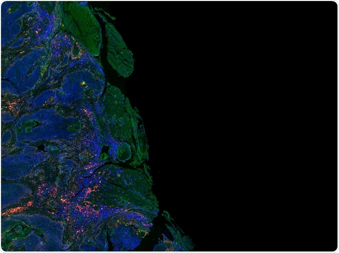
Immunofluorescence can also be applied to immunohistochemistry, however, this technique uses a fluorescent dye called a fluorophore instead of a colored substrate. The fluorophore is conjugated onto the antibody and a fluorescent microscope is used to visualize the tissue sample. Either the primary or secondary antibody can be conjugated with the fluorophore, giving rise to direct (primary antibody) or indirect (secondary antibody) immunofluorescence.
 Carl Dupont | Shutterstock
What factors affect immunohistochemistry and immunofluorescence?
Selecting an antibody
One important factor that affects immunohistochemistry and immunofluorescence is the choice of primary antibody. Cross-reactivity can pose a great limitation here. Many antibodies are available, and some may be more efficient at binding to the target protein than others. In addition, some antibodies can react with several similar proteins in a sample.
Generally, it is advisable to determine this experimentally by using the antibody to stain tissues where the target protein is known to be absent, or by staining a tissue where the localization of this protein is known.
Validation using western blotting
Western blotting could be used to determine specificity, however, it is important to note that the tertiary (3D) structure of the target protein is more likely to be preserved for an immunohistochemistry experiment compared to a Western blot.
This means that even though the antibody may be specific to the target protein in a Western blot, it may not be as specific during immunohistochemistry. It should also be noted that the way the tissue has been fixed and the way in which the target protein has been “retrieved” could also affect the specificity of the antibody.
Methodology
Factors such as the amount of antibody used, the solvent it is dissolved in, how long the antibody is mixed with the tissue for, and the temperature at which the immunohistochemistry was carried out could all affect the outcome.
For example, a long incubation time would give more time for the antibody to penetrate deeply into the tissue, which could increase the signal, while the combination of a longer incubation and a lower temperature can lead to a more specific signal.
Selecting a fluorophore
In immunofluorescence, it is also important to select an appropriate fluorophore. Factors such as excitation and emission wavelengths (spectral range) and photostability must be considered. For example, if more than one target protein is being selected, then it is better to use fluorophores with a different spectral range for the different targets.
It possible to extend the lifespan of the fluorophore through the use of certain protective reagents, as well as by limiting the exposure of these molecules to lights or by using a more protective vial.
Further Reading
Last Updated: Apr 1, 2019