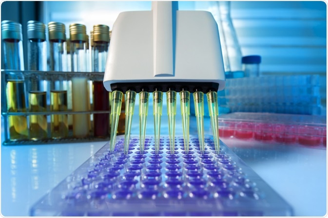
Multiple fields of microbiology rely on techniques of microbial identification, such as in food microbiology for detecting contaminants, and in microbial ecology to characterize biodiversity.
 Image Credit: Parilov / Shutterstock.com
Traditionally, microbial identification techniques have used staining, culturing, and biochemical tests to conduct phenotypic identification. However, recent years have seen progress in the methods available to conduct microbial identification, with more powerful biochemical and immunological techniques being established. Here, we discuss the method of pyrosequencing and how it is used for microbial identification.
What is pyrosequencing?
Pyrosequencing is a type of DNA sequencing that can be used for microbial genome sequencing. The technique is frequently used to identify bacterial species, distinguish bacterial strains in a sample, and identify genetic mutations that bestow anti-microbial resistance. The pyrosequencing technique has become popular within microbiology applications due to its numerous advantages, including its speed, high-throughput screening, and accuracy.
Back in 1993, scientists Mathias Uhlen and Pål Nyren first described the principle of Pyrosequencing. The method was inspired by the Sanger DNA sequencing method established in the 1970s. Pyrosequencing is a DNA sequencing method that differs from the earlier Sanger method in that instead of detecting chain termination with dideoxynucleotides, it detects the release of pyrophosphate and the generation of light on nucleotide incorporation.
The technique is fast and accurate at sequencing nucleic acids based on the "sequencing by synthesis” principle. It uses a single strand of DNA as a template and creates a complementary strand by synthesizing one base at a time.
It also detects a chemiluminescent signal to identify each incorporated base (either A, T, G, or C). Template DNA, DNA polymerase, dNTPs, ATP sulfurylase, apyrase luciferin, and luciferase are included in the pyrosequencing reaction. The addition of a dNTP as well as DNA polymerase to the template DNA initiates the synthesis reaction.
DNA polymerase is used to add the complementary dNTP onto the template, which causes pyrophosphate to be released. Next, ATP sulfurylase transforms pyrophosphate into ATP, which behaves as a catalyst, initiating the conversion of luciferin to oxyluciferin, a reaction that generates light.
The intensity of the light given off from the reaction is proportional to the number of nucleotides and can ascertain how many types of dNTPs are present within the template strand. This process is repeated, adding each of the four dNTPs separately until the entire DNA sequence of the single-stranded DNA template is established.
Using pyrosequencing for microbial identification
Below, we outline the recommended procedure for using pyrosequencing for microbial identification as put forward by Cummings et al. in 2013.
To begin with, prior to conducting the pyrosequencing process, an appropriate culture broth should be inoculated with a single bacterial colony and incubated overnight. Genomic DNA is then purified using a bacterial pellet and a genomic isolation kit. To ensure the purified genomic DNA is free of contamination from proteins it should have a 260/280 (nm) ratio >1.8. To complete the pyrosequencing process, 10-20 ng of the purified genomic DNA is required.
The next step of the process is to conduct the PCR (Polymerase chain reaction). Prior to setting up PCR, the reaction mix should be thoroughly stirred by pipetting the mixture up and down. Cummings et al. recommend that 10 ng of genomic DNA should be added into each PCR tube, and the thermal cycler should be programmed to the appropriate settings.
Finally, the PCR tubes must be placed into the cycler where the cycling program commences. The PCR process results in the amplification of the sample, which is essential for the rest of the protocol.
In the 2013 paper by Cumming et al., the following sequence of processes is recommended for successful pyrosequencing: immobilization of the biotinylated PCR products, denaturation of DNA, addition of sequencing primer, annealing of sequencing primer to DNA strands, and finally, beginning and completing the pyrosequencing run using appropriate equipment.
 Polymerase chain reaction (PCR) technique in the lab. Image Credit: angellodeco / Shutterstock.com
Analysis
Pyrosequencing software will generate data and perform various analyses. It facilitates the comparison and alignment of the sequence generated by pyrosequencing to an extensive database of bacterial ribosomal sequences. This stage of comparison and alignment allows for microbial identification.
The generated sequence can also be analyzed to detect mutations that may produce resistance to antibiotic drugs. In addition, multiple isolates taken from the same patient can be analyzed to monitor drug resistance and its progression over time to gain a deeper understanding of outbreaks of microbial drug resistance.
Overall, pyrosequencing is a rapid and reliable technique for microbial identification that can assist various fields of microbiology. It is not the only technique available for conducting microbial identification; however, it is newer than the traditional techniques and has numerous advantages, such as its accuracy.
Sources
- Ahmadian, A., Ehn, M. and Hober, S., 2006. Pyrosequencing: History, biochemistry and future. Clinica Chimica Acta, 363(1-2), pp.83-94. hackert.cm.utexas.edu/.../pyrosequencing%202006.pdf
- Cummings, P., Ahmed, R., Durocher, J., Jessen, A., Vardi, T. and Obom, K., 2013. Pyrosequencing for Microbial Identification and Characterization. Journal of Visualized Experiments, (78). https://pubmed.ncbi.nlm.nih.gov/23995536/
- Petrosino, J., Highlander, S., Luna, R., Gibbs, R. and Versalovic, J., 2009. Metagenomic Pyrosequencing and Microbial Identification. Clinical Chemistry, 55(5), pp.856-866. https://pubmed.ncbi.nlm.nih.gov/19264858/
- Tsiatis, A., Norris-Kirby, A., Rich, R., Hafez, M., Gocke, C., Eshleman, J. and Murphy, K., 2010. Comparison of Sanger Sequencing, Pyrosequencing, and Melting Curve Analysis for the Detection of KRAS Mutations. The Journal of Molecular Diagnostics, 12(4), pp.425-432. https://www.ncbi.nlm.nih.gov/pmc/articles/PMC2893626/
Further Reading
Last Updated: Jan 19, 2021