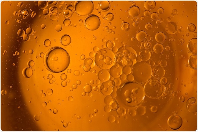
Microbubbles are small, gas-filled bubbles, typically between 0.5µm and 10µm in diameter, that are widely used as contrast agents in medical imaging and as carriers for targeted drug delivery.
 Credit: venars.original/Shutterstock.com
The core of the microbubble is a gas, which is surrounded by a shell that may be composed of polymers, lipids, lipopolymers, proteins, surfactants or a combination of these.
Microbubbles are usually injected intravenously, a process that researchers have shown is safe compared to the use of conventional contrast agents in techniques such as magnetic resonance imaging and radiography.
Development of microbubbles
Microbubbles were originally developed in the 1990s to enhance ultrasound scans. They resonate in an ultrasound beam, contracting and expanding as pressure changes occur in the ultrasound wave.
Microbubbles resonate particularly vigorously at the high frequencies used in ultrasound scans, meaning they reflect these strong waves significantly more effectively than body tissues do. Since they are approximately the same size as red blood cells, they exhibit similar rheology in blood vessels and are used to measure blood flow in organs and tumors.
Once researchers realised these bubbles could circulate through the body safely, they started to consider whether they could be used for drug delivery in the treatment of cancer.
Microbubbles in cancer
Currently, chemotherapy drugs are injected intravenously and circulate in the patient’s bloodstream. Although they are designed to destroy cancer cells, they also damage healthy tissue, leading to side effects such as nausea and hair loss.
Scientists started to use microbubbles for the targeted release of drugs, which would require significantly smaller doses than when chemotherapy drugs alone are used. The shell of the microbubble can also prevent the drug from damaging healthy cells. Initial tests have shown that using microbubbles in this way is significantly more beneficial to patients, who experience dramatically reduced side effects and recover much more rapidly.
The process involves a microbubble being loaded with a drug and antibodies that target cancer cells. The microbubbles are mixed with water and injected intravenously and tracked by ultrasound until they reach tumor tissue. The frequency of the ultrasound waves is then increased to agitate the bubbles, which burst and deliver the drug directly to the tumor cells.
The future for microbubbles
The potential medical applications of microbubbles extend beyond cancer therapy. Over the last decade, researchers have made significant advances in developing microbubbles as useful agents in molecular imaging and targeted gene delivery. Different types of microbubbles possess unique properties that can be leveraged and adjusted for specialized functions.
Microbubbles and cells interaction under high-speed microscopy
Further Reading
Last Updated: Jul 19, 2023