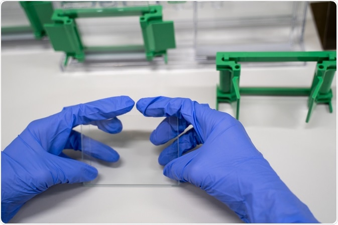
Fluorescent western blotting is a powerful protein detection technique that offers numerous benefits in comparison to alternative chemiluminescent or chromogenic detection systems. It produces less chemical waste, is faster, and is a reliable method of collecting quantitative data.
 Image Credit: ArtPanupat / Shutterstock.com
In addition, advancements in technology have increased the sensitivity of the technique, as well as reducing its cost. What’s more, the technique can be used to detect multiple proteins simultaneously, and for these reasons, Fluorescent Western blotting is rising in popularity.
How it works
In order to detect the presence of specific proteins, the fluorescent western blotting system appropriates the antigen-antibody complex, just as the alternative colorimetric and chemiluminescent western blotting methods do.
However, rather than producing signals as a result of products of enzyme-substrate reactions, as is the method of colorimetric and chemiluminescent western blotting, fluorescent western blotting, in contrast, captures signal in the form of light. A fluorophore, an inherently fluorescent molecule, produces transient light emission as it releases photons when it returns to its excited state following excitation.
The process involves separating specific proteins through electrophoresis and immobilizing them on a blotting membrane, preparing them for investigation. Detection of the protein is then achieved by adding a fluorescent dye, which is excited by exposing it to an appropriate source of light. Light at the suitable wavelength is absorbed by the dye, exciting the fluorescent molecule, releasing photons as it returns to its ground electronic state. A digital imager is then used to detect the emitted light, sensing the presence of the protein.
Optimization is also an important stage of the fluorescent western blotting system, just as it is with the alternative enzyme reaction-based methods. The degree of fluorescent labeling must be just right to generate the optimal signal level: if it is too little, then the signal is too weak, and if it is too much the signal is again too weak because of resultant inactivation of the detection reagent.
Finally, it is necessary for researchers using the fluorescent western blotting method to take special consideration of which materials they select as membranes. Those that are most common in other methods, such as nitrocellulose and polyvinylidene difluoride (PVDF), have themselves got fluorescent properties.
Using these materials in fluorescent western blotting generates background signals that impair the reading of real data. In order to address this problem, specific reagents have been created for use in this method, those that have certain excitation and emission wavelengths that reduce auto-fluorescence from common membranes. In addition, specific low-fluorescence membranes have also been created to get around this problem.
Data capture
Special fluorescence imaging equipment is required to read and document the light emitted through the fluorescent western blotting process. Fluorescence imaging equipment usually comes in the form of an illumination instrument, a laser or LED-based light source, and a filter that ensures that the correct light wavelength is delivered to the sample, as well as allowing for the capture of the necessary wavelength in the emission output.
Current instruments mostly use a charge-coupled device (CCD)–based camera to record the signal, meaning that data is available in a digital format.
As fluorescent western blotting has the capability of multiplex detection, allowing for multiple targets to be identified simultaneously, further tools are necessary to analyze the multiple fluorescent dyes used to detect distinct targets. Most commonly, online tools are employed to aid panel selection for multiplex fluorescence detection.
Benefits of fluorescent western blot detection
The method of fluorescent western blot detection has several advantages over the alternative, enzyme-based detection methods. These benefits are helping to establish it as an increasingly popular method of protein detection.
As discussed, the detection of multiple proteins is easily achievable with the fluorescent western blot method. From just one single blot, multiple proteins can be detected at the same time using spectrally distinct dyes that emit light at signature wavelengths, allowing for multiple targets to be registered without the need for separate testing.
The method is also considered to be successful at providing reliable quantitative data. It has been seen that the levels of signal produced are proportional to the volume of protein in the sample. Proteins that are both in abundance, or only sparsely dispersed in a sample can be accurately identified, giving the method power in quantitative detection. In comparison with the alternative enzyme systems, fluorescent western blotting can result in more quantitative data.
Also, the method has the advantage of saving researchers time. It can do this in multiple ways. Firstly, because it can detect numerous target proteins from just one blot sample, rather than conducting multiple tests. Secondly, because with this kind of testing, there is no need to allocate time to substrate incubation or film exposures.
Further to this, the use of particular fluorescent dyes has been proven to be high in sensitivity, leading to a robust method of protein detection. Because of their low background autofluorescence, low fluorescence quenching, and high extinction coefficients, far-red and infrared fluorescent dye conjugates result in greater detection sensitivity.
Overall, fluorescent western blotting has established itself as a robust method of protein detection as it offers numerous benefits over alternative methods, such as the ability to detect multiple proteins simultaneously, its high sensitivity, and time-saving processes. The result is that it is becoming an increasingly popular technique for protein detection.
Sources:
- Eaton, S., Roche, S., Llavero Hurtado, M., Oldknow, K., Farquharson, C., Gillingwater, T., and Wishart, T. (2013). Total Protein Analysis as a Reliable Loading Control for Quantitative Fluorescent Western Blotting. PLoS ONE, 8(8), p.e72457. https://journals.plos.org/plosone/article?id=10.1371/journal.pone.0072457
- Jacksonimmuno.com. (2019). [online] Available at: https://jacksonimmuno.com/secondary-antibody-resource/immuno-techniques/fluorescent-western-blotting/?pdf=126 [Accessed 17 Oct. 2019].
- Towbin, H., Staehelin, T., and Gordon, J. (1979). Electrophoretic transfer of proteins from polyacrylamide gels to nitrocellulose sheets: procedure and some applications. Proceedings of the National Academy of Sciences, 76(9), pp.4350-4354. https://www.ncbi.nlm.nih.gov/pubmed/388439
- Ni, D., Xu, P., and Gallagher, S. (2016). Immunoblotting and Immunodetection. Current Protocols in Immunology, 114(1), pp.8.10.1-8.10.36. https://currentprotocols.onlinelibrary.wiley.com/doi/abs/10.1002/cpim.10
Further Reading
Last Updated: Dec 12, 2019