Aug 16 2011
Until now, brain scans obtained using fMRI – one of the most important imaging methods in use today – have been affected by fluctuations in the brain activity measured which could not be explained. Researchers at the Max Planck Institute for Human Cognitive and Brain Sciences in Leipzig and the Charité in Berlin have now shown that certain electric background rhythms in the brain, known as Alpha oscillations, alter the fMRI signal obtained with visual stimuli. To do this, they used a new approach in which quickly changing brain activity, which cannot be captured by the scanner, was measured using
Electroencephalography (EEG). This showed that the strength of the oscillations also influenced the reaction times of the study participants. These new findings could help optimise fMRI and also offer new approaches for targeted therapeutic manipulation of brain activity.

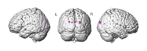
Purple regions indicate sites of interaction between brain electric oscillations and fMRI responses to optical stimuli
No other method has influenced brain research like fMRI. Most people are familiar with the images showing areas of changed brain activity in different colours. Its good spacial resolution and the fact that images can be obtained without the need for radiation have made fMRI the method of choice for brain imaging. However, fMRI also has disadvantages. In addition to the brain signal, the scanners also pick up a great deal of “noise” – signals which are unrelated to actual cognition. It can happen that greatly differing brain activity is measured in the same person and the same task. It is difficult to decide whether the fluctuations in the signal reflect actual cognitive reactions in the brain or other noise. This has frequently led to criticism of the technique and the images it produces.
“fMRI data normally reflect several processes happening at the same time” says Petra Ritter, Group Leader of the Minerva Research Group ‘Brain Modes’ at the Max Planck Institute for Human Cognitive and Brain Sciences in Leipzig. Technical factors as well as factors like heartbeat and breathing can distort the data, explains the scientist, who is also Coordinator of the Bernstein Focus ‘State Dependencies of Learning’ and conducts research at the Charité in Berlin. Because fMRI scans can be misinterpreted, it is important to apply appropriate methods to filter out sources of data distortion as accurately as possible.
Ritter and her team of researchers have been investigating a new approach, the simultaneous use of fMRI and EEG, which aims to illuminate this issue. Using EEG, the scientists identify the fluctuating electrical brain oscillations in real time – that is during fMRI scanning and not after it, as is typical. By doing this, Ritter, together with post-doctoral researcher Robert Becker and a team of scientists were able to answer a previously unanswered question: Do – in addition to the non-neuronal noise – fluctuations in electrical background activity alter the fMRI signal also? The team of researchers examined the influence of Alpha oscillations to do this. The Alpha rhythm is the temporal pattern in which the neurons in the visual system oscillate when a person is in a relaxed waking state. Whenever the EEG of the person in the scanner showed strong Alpha activity, an optical stimulus was shown. The results demonstrated that the Alpha oscillations systematically changed the fMRI response to the stimulus and therefore were also responsible for its variability. In addition, the strength of the Alpha oscillations influenced the speed of the participants’ reactions to the stimuli.
“This knowledge is important for understanding the neuronal mechanisms that are the foundation of variation in brain signals and, consequently, contribute to variations in behaviour”, says Becker. This explains why subjects can show changing responses in brain activity or even behavior, even if the experimental set-up is kept exactly the same. The results also demonstrate the usefulness of the method of combining fMRI and EEG, a method which has the potential to be used in real-time monitoring of background brain activity and in targeted manipulations which could be used in therapeutic alterations of behaviour.
fMRI/EEG
Simultaneous fMRI/EEG acquisition is not easy as the magnetic field of the scanner influences the EEG signal. It is worth solving this issue, however, as both methods complement each other. fMRI can provide very exact images of the location of changes in blood flow in the brain, but these do not reflect the time course of these changes. EEG, in contrast, measures electrical activity of nerve cells directly and with millisecond precision, but the source of these changes can only be very approximately located.
Original publication:
Becker R, Reinacher M, Freyer F, Villringer A, Ritter P. 2011 How ongoing neuronal oscillations account for variability of evoked fMRI responses. J Neuroscience 31(30), S. 11016-27.
Contact:
Dr. Robert Becker
Charité, University Medicine Berlin
Email: [email protected]
PD Dr. Petra Ritter
Max Planck Institute for Human Cognitive and Brain Sciences/ Charité, University Medicine Berlin
Phone.: +49 30 450 560 005
Email: [email protected] or [email protected]
Peter Zekert
Max Planck Institute for Human Cognitive and Brain Sciences Press and Public Relations Phone: +49 341 9940-2404 Email: [email protected]