May 12 2016
LaVison BioTec, developers of advanced microscopy solutions for the life sciences, report on the work of Nicolas Renier, a Post-Doctoral Fellow in the laboratory of Marc Tessier-Lavigne at the Rockefeller University in New York where he applies light sheet microscopy to study axon rewiring in adult nervous systems.
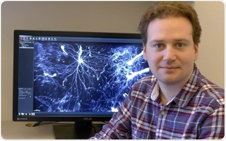
Dr Nicolas Renier from the Rockefeller University, NY, with results from his LaVision BioTec UltraMicroscope light sheet microscope.
Drs Nicolas Renier and Zhuhao Wu are post-doctoral fellows in the laboratory of Marc Tessier-Lavigne at the Rockefeller University in New York where they have co-developed methodologies to develop new imaging techniques applying light sheet microscopy to the study axon rewiring in adult nervous systems. It is hoped that this could lead to an understanding as to how experience or even pathologies such as Alzheimer’s disease modify the way the brain is wired.
Two great advantages have been behind their motivation to use the LaVision BioTec UltraMicroscope: high speed and large fields of view. For neuroanatomy, most of the time, users want to look at large regions, up to 1 cm wide. Also, there is the need to image a lot of samples to get statistical significance, so a very high acquisition speed is required. To date, they feel this is the only microscope on the market that can do that so far.
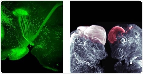
Examples of the stunning image quality generated By Dr Nicolas Renier using his UltraMicroscope light sheet microscope.
2-photon microscopy is sometimes used for volume imaging when the need is to go to higher magnifications, and confocal or epifluorescence microscopies are used for classical histology done on sections. Talking about his use of the UltraMicroscope, Dr Renier says:
This is the only microscope that has a built-in compatibility with the 3DISCO clearing method, which in our hands is still the best clearing method. The main strength of the technique is the ease of implementation and flexibility in sample handling. The speed also compares favorably to other methods. Lastly, the signal to noise ratio of Alexa dyes combined with 3DISCO clearing and light sheet microscopy is extremely high.
Asked how he sees the technique and methodology develop, Dr Renier says:
We are now using iDISCO routinely in the lab for most of our projects. The technique has upgraded most of our histology assays and we now rarely section tissues anymore. We use the technique to study the trajectory of nerves in whole embryos, to count cell number in whole organs, to trace single axons in the brain. The technique can be used for large screens, thus enabling quick phenotyping of mutants, something that is usually challenging in mice. We are working on trying to take the technique beyond classical immunostaining. Volume mRNA in situ hybridization is an avenue to be explored. We are also looking at testing additional labeling techniques, including the use of llama nanobodies and chemical tag based labeling.
 New research decodes the bacterial “zip code” of colorectal cancer for prediction and survival