Sep 11 2017
Researchers at the Helmholtz Zentrum München have developed a new method for reconstructing continuous biological processes, such as disease progression, using image data. The study was published in ‘Nature Communications’.
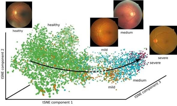
The new method is able to reconstruct biological processes using image data. Source: Helmholtz Zentrum München
Modern life sciences generate a constantly growing amount of data in shorter and shorter cycles. Making such data controllable and suitable for evaluation is the objective of Dr. Alexander Wolf and his colleagues at the Helmholtz Zentrum München’s Institute of Computational Biology (ICB). With this in mind, the researchers are attempting to develop software that handles this evaluation. But of course there are various hurdles to clear.
“In the current study, we dealt with the problem that software cannot assign image data to continuous processes,” explains study leader Wolf. “For example, it is possible to classify image information according to clearly defined categories, but in disease progression and developmental biology, the limits are quickly reached because the processes are continuous and not individual steps.”
In order to take this into account, the Helmholtz team employed methods from so-called Deep Learning (i.e. machine learning processes). “Using artificial neural networks, we can now combine individual pictures into processes and additionally display them in a way that humans understand,” say Philipp Eulenberg and Niklas Köhler, former Master’s students at the ICB and the study’s first authors.
Blood cells and retinas as sparring partners
In order to demonstrate the method’s capability, the scientists selected two examples. In the first approach, the software reconstructed the continuous cell cycle of white blood cells using images from an imaging flow cytometer (producing pictures in a fluorescence microscope). “A further advantage of this examination is that our software is so fast that it is possible to extract the cell development on the fly, meaning while the analysis in the cytometer is still running,” explains Wolf. “In addition, our software makes six times less errors than previous approaches.”
In the second experiment, the researchers reconstructed the progress of diabetic retinopathy. “We did this by feeding our software 30,000 individual images of retinas as sparring partners, so to speak,” explains Niklas Köhler. “Since it automatically compiles these data into a continuous process, the software allows us to predict the disease progression on a continuous scale.”
And if the data are not part of a continuous biological process? “In such a case, the software recognizes that individual categories are involved and assigns the measured data to individual clusters,” Wolf explains. In addition to further applications for the method, in the future Wolf and his colleagues want to solve other problems involving the evaluation of biological data using machine learning.
Source: https://www.helmholtz-muenchen.de/en/news/latest-news/press-information-news/article/41458/index.html
 New partnership aims to expand global access to sickle cell gene therapy