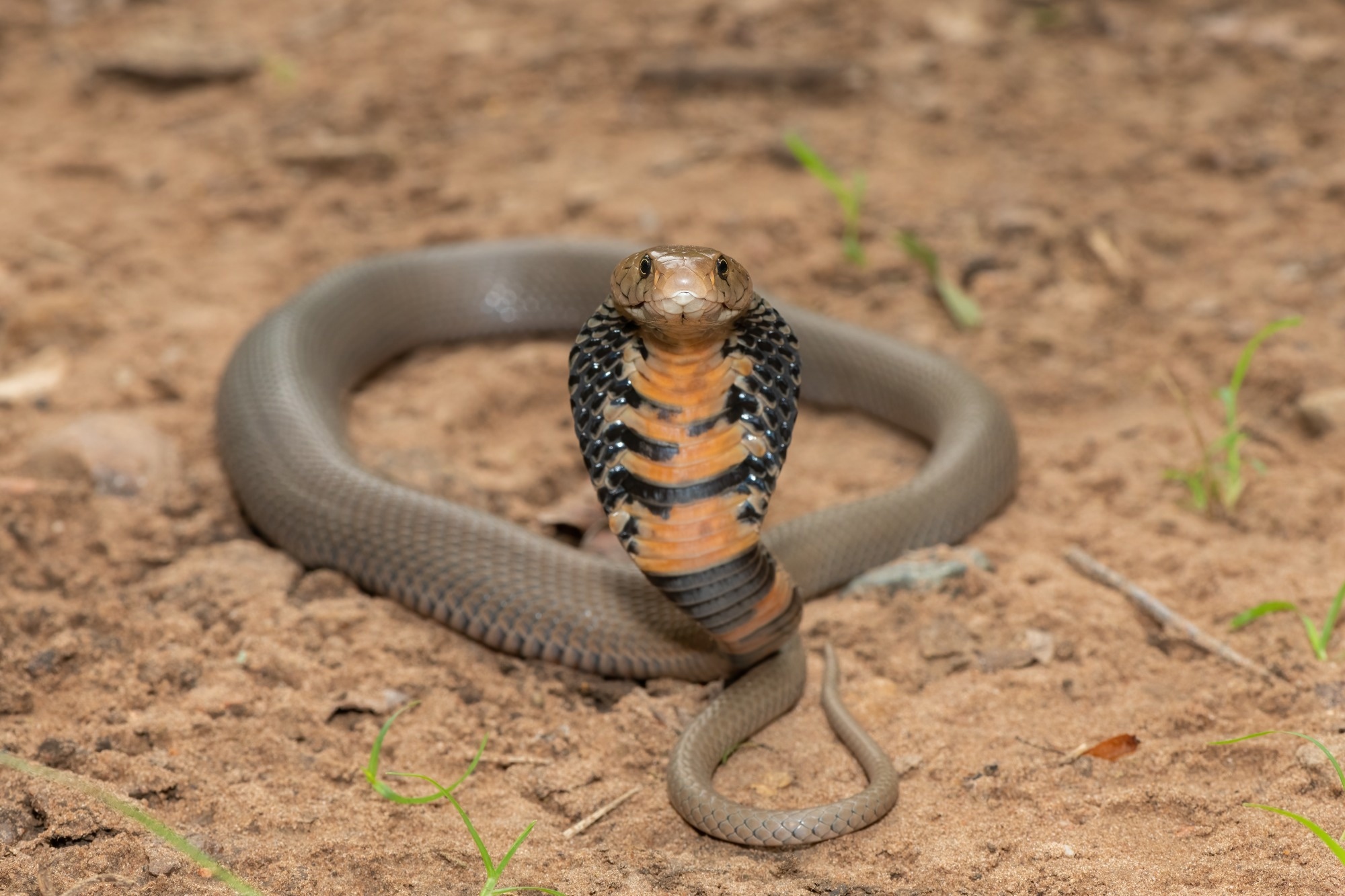
Snakebite is a neglected tropical disease, mainly affecting rural communities in Latin America, South Asia, Southeast Asia, and sub-Saharan Africa. An estimated 138,000 deaths occur due to snakebites annually. Severe local pathology is often observed at the bite site after viper envenoming, which results from hemorrhagic, cytotoxic, or myotoxic venom toxins. Envenoming by elapid snakes leads to neurotoxic muscle paralysis with no tissue damage.
However, envenoming by cobras, mainly African spitting cobras, causes little neurotoxicity but rapid and severe swelling and tissue destruction, often leading to necrosis. Cytotoxic three-finger toxins (CTx) are the most abundant in many cobra venoms and are associated with dermonecrotic pathology. CTx can disrupt cell membranes and induce pore formation, leading to cell death. Phospholipases A2 (PLA2) are the second most abundant in several cobra venoms.
Intravenous antivenom therapy is the current treatment for snakebite envenoming. However, several limitations restrict its clinical utility, such as limited efficacy due to toxin variations, low affordability, and severe adverse reactions. Further, treatment initiation in a clinical setting is delayed due to poor hospital accessibility in rural tropical areas where snakebites are prevalent. Thus, new therapies are needed to overcome these limitations.
 Study: Dermonecrosis caused by a spitting cobra snakebite results from toxin potentiation and is prevented by the repurposed drug varespladib. Image Credit: Craig Cordier / Shutterstock
The study and findings
In the present study, researchers identified the venom toxins responsible for dermonecrosis caused by African spitting cobras. First, mice were intradermally challenged with African spitting cobra venom. Mice received doses from the West and East African forms of Naja nigricollis, the black-necked spitting cobra. The resultant dermonecrotic lesions were analyzed 72 hours later.
The lesions were heterogeneous, with a white region of tissue damage surrounding the dark necrotic center. Further, hematoxylin and eosin staining revealed conspicuous histological differences between macroscopically dark and white areas. Samples from the dark lesions showed extensive damage to all layers of the skin; the epidermis was lost, while the hypodermis and dermis were severely damaged.
Contrastingly, white lesions showed an inflammatory infiltrate in the dermis and hyperplasia of the epidermis. Next, the team quantified the cytotoxic potency of the venom constituents using the immortalized human epidermal keratinocytes. Cells were exposed to crude venom, two purified PLA2s (acidic and basic), four purified CTx (CTx1, CTx1v, CTx3, and CTx4), or combinations. Crude venom significantly reduced cell viability.
![African spitting cobra venoms cause heterogenous dermonecrotic lesions in vivo. Groups of mice (n = 3) were injected intradermally with two doses of spitting cobra venom and after 72 h the resulting lesions were excised for macroscopic quantification of damaged areas and histological assessment. (A) Representative macroscopic image of a skin lesion induced by 100 µg of venom from West African (Nigeria) N. nigricollis, in which a dark central area (D) of necrosis is observed surrounded by a white area (W) of skin damage. (B–D) Representative light micrographs of sections of the skin of mice injected with PBS or West African N. nigricollis venom. (B) Skin injected with PBS showed a normal histological appearance including the epidermis (E), dermis (D), hypodermis (H), panniculus carnosus (P) and adventitia (A). (C) Light micrograph of a section of skin corresponding to a dark area of venom-induced damage. All skin layers were affected, with loss of epidermis (arrow) and skin appendages in the dermis. A proteinaceous hyaline material was observed (*). (D) Light micrograph of a section of the skin corresponding to a white area of damage from a mouse injected with venom. There was an increase in the thickness of epidermis (hyperplasia; arrow) and inflammatory infiltrate in the dermis. Thrombi (T) were observed in some blood vessels. (E) The area of dermonecrotic lesions caused by N. nigricollis (West African, Nigeria [NGA]; East African, Tanzania [TZA]) venoms at different doses. Bars show the mean area of the total lesions (T) in comparison to the dark central areas (D) of greatest intensity, and error bars represent the SD from the mean. Scale bar in (B–D) represent 100 µm.](https://www.news-medical.net/images/news/ImageForNews_779485_17153110398505466.jpg)
African spitting cobra venoms cause heterogenous dermonecrotic lesions in vivo. Groups of mice (n = 3) were injected intradermally with two doses of spitting cobra venom and after 72 h the resulting lesions were excised for macroscopic quantification of damaged areas and histological assessment. (A) Representative macroscopic image of a skin lesion induced by 100 µg of venom from West African (Nigeria) N. nigricollis, in which a dark central area (D) of necrosis is observed surrounded by a white area (W) of skin damage. (B–D) Representative light micrographs of sections of the skin of mice injected with PBS or West African N. nigricollis venom. (B) Skin injected with PBS showed a normal histological appearance including the epidermis (E), dermis (D), hypodermis (H), panniculus carnosus (P) and adventitia (A). (C) Light micrograph of a section of skin corresponding to a dark area of venom-induced damage. All skin layers were affected, with loss of epidermis (arrow) and skin appendages in the dermis. A proteinaceous hyaline material was observed (*). (D) Light micrograph of a section of the skin corresponding to a white area of damage from a mouse injected with venom. There was an increase in the thickness of epidermis (hyperplasia; arrow) and inflammatory infiltrate in the dermis. Thrombi (T) were observed in some blood vessels. (E) The area of dermonecrotic lesions caused by N. nigricollis (West African, Nigeria [NGA]; East African, Tanzania [TZA]) venoms at different doses. Bars show the mean area of the total lesions (T) in comparison to the dark central areas (D) of greatest intensity, and error bars represent the SD from the mean. Scale bar in (B–D) represent 100 µm.
While all four CTx reduced cell viability, CTx3 was the most potent. The two PLA2s alone or in combination were not sufficiently toxic. Additional experiments revealed that mice receiving the CTx plus PLA2 combinations had developed lesions similar in size to those induced by crude venom; this suggested that these two toxin groups recapitulate most of the crude venom effects.
The team speculated that inhibiting one of the two groups may reduce venom-induced dermonecrosis. As such, they repeated the experiments using varespladib, a PLA2 inhibitor. Varespladib was preincubated with the venom or purified constituents before the intradermal challenge. The co-injection of varespladib and crude venom significantly decreased lesions from 52 mm2 to 2.6 mm2. Likewise, varespladib and CTx plus PLA2 combination reduced lesions to 5.5 mm2.
Further, to mimic a realistic scenario, mice were intradermally injected with the venom, followed by varespladib injection at 0, 15, or 60 minutes later at the same site. There was a significant decline in lesion sizes compared to venom-only controls in all conditions. However, the most substantial reduction occurred when the inhibitor was injected immediately after the venom challenge.
Although the therapeutic potency decreased with longer delays between venom and varespladib injections, lesion size reductions were still significant. Notably, when varespladib was administered intravenously, no significant reductions in lesion sizes were observed, even when injected immediately after the challenge. Finally, the team investigated the effects of varespladib on venom-induced myotoxicity. Myotoxicity was induced by injecting the venom intramuscularly.
Muscle damage was assessed by quantifying plasma creatine kinase (CK) activity three hours later. The researchers noted significant reductions in plasma CK levels when varespladib and venom were co-injected. They also explored whether this efficacy was retained with delayed administration of varespladib through intravenous and intramuscular routes. This showed significant reductions in plasma CK levels via both routes, regardless of immediate or delayed treatment.
Conclusions
In sum, the study characterized the pathology of dermonecrosis caused by N. nigricollis venom in mice. Although CTx was predominantly responsible for in vitro cytotoxicity, the combinations of CTx and PLA2 were necessary to replicate the cytotoxic potency of the crude venom. Further, the team demonstrated that varespladib co-injection inhibited lesion formation; besides, varespladib did not inhibit CTx activity, supporting that using inhibitors against a single toxin family could reduce local envenoming severity.
Journal reference:
- Bartlett KE, Hall SR, Rasmussen SA, et al. Dermonecrosis caused by a spitting cobra snakebite results from toxin potentiation and is prevented by the repurposed drug varespladib. Proc Natl Acad Sci USA, 2024, DOI: 10.1073/pnas.2315597121, https://www.pnas.org/doi/full/10.1073/pnas.2315597121
 Everyday PFAS exposure alters placental function in early pregnancy