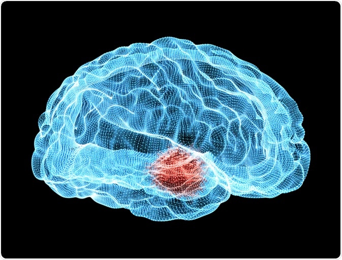
Alpha-synuclein proteins have been implicated in several neuropathological diseases.

Image Credit: Naeblys/Shutterstock.com
The de-regulation of the protein is thought to influence Parkinson’s disease, dementia, and other illnesses through the build-up of its intraneuronal aggregates, known as Lewy bodies.
Research using active alpha-synuclein proteins has proven invaluable in understanding more about the role of Lewy bodies in pathology, along with assisting in the development of new pharmaceutical drugs for the treatment of Parkinson’s disease, Lewy body dementia, and other disorders.
Alpha-Synuclein’s role in disease
The human heart, gut, muscles, and brain are all home to the alpha-synuclein (α-syn) protein. While its functions are not 100% clear, there is a vast body of evidence that has implicated it as being fundamental to numerous key processes, most notably it appears to be essential for dopamine regulation.
Studies have shown that the protein is responsible for managing the process of transporting neurochemicals between synapses, known as neurotransmission.
Dopamine plays a fundamental role in the initiation and development of Parkinson’s disease (PD), and research has shown that there is a link between alpha-synuclein and the disease that looks likely to have a genetic basis. In addition to this, it has been found that alpha-synuclein is involved in multiple system atrophy (MSA), dementia with Lewy bodies (DLB), and pure autonomic failure (PAF).
Along with Parkinson’s disease, these diseases are known as the synucleinopathies, due to their close link with alpha-synuclein. Evidence has uncovered that pathological deposition of these protein aggregates, or Lewy bodies (LBs), are linked with the synucleinopathies.
Unsurprisingly, much research has been conducted in this area to understand the cause and development of these disorders, as well as to explore potential new therapeutic avenues.
Lewy bodies are aggregates of alpha-synuclein
The alpha-synuclein protein aggregates in neuropathological diseases, such as Parkinson’s. The formation of these toxic clumps is characteristic of the illness, and they are associated with the related death of brain cells.
For this reason, the de-regulation of the alpha-synuclein protein is considered to be an underlying cause of the synucleinopathies. Tests to identify these protein clumps or Lewy bodies is essential to early, accurate diagnosis because often symptoms of certain synucleinopathies resemble those of Alzheimer's or even psychiatric illnesses.
Testing for Lewy bodies, the intraneuronal aggregates of the alpha-synuclein protein, is therefore essential to the continued research of Parkinson’s disease, Lewy body dementia, and other disorders.
Research techniques with active alpha-synuclein proteins
Experimentally, active alpha-synuclein proteins can be used to induce Lewy bodies so that they can be studied in specific, controlled scenarios. These kinds of studies can elucidate the behavior of Lewy bodies, and uncover more about their role in various diseases.
Methods have also been developed to detect endogenous active alpha-synuclein protein levels in a sample using a dye.
These techniques can be used to study samples both in vivo and in vitro, and they can look into not only the functions of Lewy bodies in pathology but can also assist in the development of new pharmaceutical drugs through testing their impact on Lewy bodies.
Fluorescence techniques are the most common method for identifying active alpha-synuclein proteins. Thioflavin T (ThT) is one of the most popular dyes used in this assay. The process involves the seeding of new alpha-synuclein aggregates from a pool of active alpha-synuclein monomers.
The fluorescent ThT dye is added to bind to the beta sheet-rich structures that are found in alpha-synuclein aggregates. The event of ThT binding with the structures in the aggregates causes a shift in the dye’s emission spectrum, turning it red and increasing its fluorescence intensity.
Past research has used this technique to identify the aggregation of these endogenous proteins in Alzheimer’s disease (AD), Parkinson’s disease, amyloidosis, and others. The methods can be constructed also in vitro to allow for the study of interactions in the absence of other influencing factors.
Modern studies mostly perform assays with Thioflavin T within fluorescence plate readers, which allow for multiple conditions to be explored at the same time. While it is an established technique that is frequently used for looking at active alpha-synuclein and its aggregates, the process is not without its limitations.
The method is known for its poor reproducibility, mostly due to the number of varying factors that affect protein aggregation. Currently, scientists are working on improving this method to increase the reproducibility of ThT assays. Recent developments of the method have involved adding a period of orbital shaking, as well as improving mixing with the addition of glass beads.
Future directions
Active alpha-synuclein proteins provide a robust method for looking at the Lewy bodies associated with numerous neuropathological diseases. They are currently being used to understand the impact of alpha-synuclein on these diseases, as well as to develop new treatment methods.
The most common method of investigating them is through fluorescence techniques that use Thioflavin T as a dye. While this method is reliable and well established, scientists are currently working on improving its reproducibility to enhance the strength and accuracy of the method.
Sources:
Li, X., Dong, C., Hoffmann, M., Garen, C., Cortez, L., Petersen, N. and Woodside, M. (2019). Early stages of aggregation of engineered α-synuclein monomers and oligomers in solution. Scientific Reports, 9(1). https://www.nature.com/articles/s41598-018-37584-6
Narkiewicz, J., Giachin, G. and Legname, G. (2014). In vitro aggregation assays for the characterization of α-synuclein prion-like properties. Prion, 8(1), pp.19-32. https://www.ncbi.nlm.nih.gov/pmc/articles/PMC6066150/
Wördehoff, M. and Hoyer, W. (2018). α-Synuclein Aggregation Monitored by Thioflavin T Fluorescence Assay. BIO-PROTOCOL, 8(14). https://www.ncbi.nlm.nih.gov/pmc/articles/PMC6066150/
Further Reading
Last Updated: Feb 3, 2020