Necroptosis is a newly characterized form of cell death that has similar properties to both apoptosis and necrosis. Necroptosis has been associated with various disease states, therefore research and analysis of cells undergoing this cell death pathway is vital.
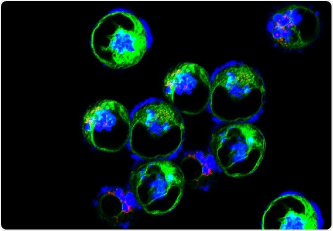 Image Credit: Vshivkova / Shutterstock
What is necroptosis?
Necroptosis is a form of programmed cell death, distinguished by rupturing of the cell membrane, swelling of the organelles, and spillage of internal cellular components. This form of cell death occurs when apoptosis cannot be carried out, for example when caspase 8 protease is inhibited by the vaccinia virus.
The pathway of this process has been extensively characterized and is highly controlled. Overall, tumor necrosis factor alpha (TNFα) signaling results in activation of receptor interacting kinase (RIPK) 1, recruitment of RIPK 3, formation of the necrosome, and the phosphorylation and oligomerization of mixed lineage kinase domain-like (MLKL). This activated form of MLKL ultimately inserts into the cell membrane and leads to permeabilization and death of the cell.
This pathway is very important in research as it has recently been suggested as a possible area of interest for cancer treatment. One of the main hallmarks of cancer is the evasion of apoptosis, which results in immortal cells that evade cellular death. Therefore, it has recently been suggested that activation of necroptosis in cancer-specific cells could help to overcome this resistance.
There are also many diseases in which necroptosis is implicated, including stroke and myocardial infarction. Therefore, it is very important that researchers can analyze and characterize cells undergoing necroptosis. Such endeavors have led to several different mechanisms for studying them, as described below.
Detecting the presence of necroptosis via biomarkers
A biomarker is a substance which is present in a living organism that can be quantified to analyze a biological process. There are many different biomarkers which have been utilized to indicate the presence of necroptosis in both in vitro and in vivo studies.
For example, several in vitro studies identified RIPK1/3 complex through immunoprecipitation and electron microscopy analysis, which indicates the presence of the necrosome and, in turn, the presence of necroptosis.
Additionally, activation of RIPK1 can be detected through western blotting against phosphorylated hRIP1 at S166. Thirdly, activation of RIPK3 can be identified through the western blot detection of phosphorylated mRTP3 at S232. Another method frequently used is through the detection of caspase 8 activity, which suggests that necroptosis is not occurring.
However, the most accurate and, therefore, most important methods of in vitro necroptosis analysis are measurements of MKLK phosphorylation, oligomerization or translocation. This can be carried out via detection of various biomarkers – Including phosphorylated MLKL at T357/S358 through western blotting or immunoprecipitation, detection of oligomerized MLKL through western blotting or through detection of MLKL localization through immunostaining, respectively.
Furthermore, there are also immunohistochemistry biomarkers which have been used in vivo to identify necroptosis in clinical studies and animal models (for example, detection of phosphorylated hMLKL at Thr357 and Ser358). This can demonstrate that necroptosis is occurring in tissue samples, which particularly aids research into diseases where necroptosis is implicated. For example, a recent study has shown that necroptosis is activated in cortical lesions of patients with multiple sclerosis.
The use of necroptosis inhibitors for analysis purposes
Another way of analyzing necroptosis is through inhibiting the process, revealing whether the specific form of cell death implicated in disease is necroptosis.
There are several different types of inhibitors frequently used. These include inhibitors of RIPK1, such as Necrostatin-1 and 7-Cl-O-Nec-1. This method, however, is not the most reliable since Nec1 has off target activity and RIPK1 is also involved in apoptosis.
Other inhibitors frequently used in analysis of necroptosis include GSK-872, which inhibits RIPK3, and necrosulfonamide, which inhibits MLKL.
Further Reading
Last Updated: Jul 19, 2023
 Study identifies PUF60 as a critical vulnerability in triple negative breast cancer