If junk is not removed, pathological conditions can develop. For example, in one condition, the neutrophil count significantly decreases. Neutrophils remove pathogens and people with a reduced neutrophil count are more prone to infection, especially to rare bacteria that wouldn’t cause infection under normal conditions.
We also grow junk in the body as cells die and get recycled. Dying cells need to be removed because they are very toxic to the surrounding environment. The cells contain dangerous biomolecules such as enzymes that cleave proteins, DNA and RNA.
How are these undesirables usually removed from the body?
Neutrophils and macrophages are the two main garbage collectors. These are referred to as phagocytes, cells that eat other components.
Neutrophils mostly target outside enemies such as pathogens and bacteria, while macrophages generally target the internal junk such as dying cells and senescent cells.
Both neutrophils and macrophages use a similar strategy to remove those undesirables. First, they move to the site where the undesirables are. Usually, these undesirables release chemicals that the neutrophils and macrophages use as a cue to locate their sites.
Once they reach their target sites, the neutrophils and macrophages communicate with the undesirables, to confirm they are definitely to be removed. They then start to activate the internal cytoskeleton which is contains the actin machinery. This physically distorts the membranes of macrophages and neutrophils, which helps to engulf or ingest the undesirables.
Next, the ingested undesirables fuse to lysosomes, which are small organelles within cells that contain digestive enzymes. They break down the undesirables so that the dying cells’ toxic contents will not be released into the environment.
So is the process of phagocytosis limited by the number of macrophages and neutrophils?
I think the answer is yes, because when a person’s neutrophil count is depleted, they are very prone to bacterial infection. A decreasing neutrophil count is strongly correlated with a decrease in the defence mechanism.
Why can’t other types of cell perform phagocytosis and what are the minimum tools a cell needs to eat another?
Phagocytosis is a very elaborate process - the initial chemical release to locate the undesirables, followed by the eating process. These processes require at least dozens, if not hundreds, of signalling components. You need to have the right set of molecules at the right time.
Neutrophils and macrophages are highly differentiated to perform phagocytosis. Both of these white blood cells originate from the same stem cells, but then differentiate into highly specialized individual cell types.
Aside from retinal pigment epithelium, most other cell types in the body are not capable of phagocytosis. They have the genetic components needed, but they are not capable of turning those genes on at the right time.
Please can you outline your research that involved looking at normally inert cells and trying to make them able to recognize and engulf dying cells?
That was a combination of new and old techniques. The new technique is what we developed and published in our present work and it is called Dimerization-Induced Surface Display (DISplay). Basically, we developed a technique to rapidly and inducibly display molecules of interest at the cell surface. This allows us to present the molecules that recognize the undesirables.
The old technique is to activate the actin machinery to induce membrane deformation. We introduce a genetically mutated molecule called Rac, which is always active, so that the membrane deformation is enhanced.
By combining these two techniques, we can make cells phagocytic. Inert cells that could not otherwise perform phagocytosis can be induced to perform phagocytosis.
What were the main challenges you faced?
We faced two challenges. During the development of the DISplay technique, we had hypothesised how things would work, but the initial trial didn’t work out.
The DISplay technique requires two components: one in the plasma membrane and another one in the Golgi complex. We made different versions of these two components and we tried to combine them so that we could find which one met our standard. That was the first challenge.
The second challenge concerned the logical application. Once we established the DISplay, we wanted to use that for phagocytosis. The initial idea was to make healthy cells mimic dying cells so that they could be eaten by macrophages.
At the time, we wanted to find out what the minimal signalling required was for the cells to be recognized and eaten by macrophages. That didn’t work out and we still haven’t figured it out.
We presented an ‘eat me’ signal because dying cells usually present an ‘eat me’ signal on their surface that is recognized by macrophages. However, displaying that signal somehow didn’t work out. We then changed our way of thinking and added the cells that can eat the dying cells.
Were the engulfed cells broken down like in natural phagocytosis?
We are interested in whether the cells that are artificially engulfed degrade afterwards or not. However, it was difficult to tell if the engulfed cells were alive or dead. They were already apoptotic, so I would say they were dying, but we don’t know whether they underwent degradation afterwards.
It was experimentally challenging. We actually expect that they are not degraded, because we didn’t turn on the lysosomal fusion process. That process requires a protein called Rab5 and we didn’t turn that signalling pathway on. We don’t think the engulfed cells were being broken down but we didn’t have direct evidence.
What impact do you think this study will have?
There are two major impacts. One is associated with the DISplay technique. We’re proud of this technique because it has fast kinetics, meaning we can present a protein of interest on the cell surface within one hour.
The cell surface is critical for cells interacting with the environment, which includes other cells and the extracellular matrix that supports those cells. Most of the information comes from outside the cell and the information process is initiated at the cell surface. Now we have a way to manipulate the information source in a rapidly inducible manner, which is a very significant achievement.
The second impact concerns the artificial phagocytosis. We tried to mimic what macrophages do in the body and what we are doing with the system is a good foundation for further engineering. We hope that the degradation would be much more efficient than the natural process and think we may be able to create super phagocytes.
We may also be able to target cells or junk other than the dying cells, such as cancer cells. If we could engineer artificial phagocytes that can target cancer cells or the amyloid-beta plaques that cause Alzheimer’s disease, for example, that could be very influential in a clinical setting.
What are the next steps in your research?
We would like to complete the artificial phagocytosis and degradation of engulfed cells, but this requires the development of a new technique.
Rab5 is responsible for the lysosomal fusion event, and we have a very good idea of how to turn this molecule on, but we would prefer to simplify this and directly fuse the engulfed cells.
Engulfed cells are wrapped up by the membrane, so we’d like to fuse this membrane to the lysosome so that the engulfed cell can be broken down.
What are the potential future implications of the ability to use artificial cells to recognize and engulf dying cells?
The artificial phagocytosis may be more efficient than the natural process and we may also be able to target other types of pathological cells, such as cancer cells. To achieve that, we are wondering about using the DISplay technique to present the antibody that recognizes the signature molecules on the surface of cancer cells.
For our DISplay technique, we need to make the DNA that fuses our molecular components with the antibody. Antibodies are very challenging to encode but there’s an antibody that’s made of a single chain. There are particular animal species that produce those single-chain antibodies and they can be encoded by the DNA plasmid. We are investigating this to see if we can target the cancer cells, amyloid-beta, and other undesirables in our bodies.
Where can readers find more information?
For information, please visit: https://www.jhu.edu/
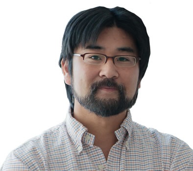
About Dr. Takanari Inoue
 Dr. Takanari Inoue is an associate professor of cell biology at the Johns Hopkins University School of Medicine. His research focuses on synthetic cell biology to dissect and reconstitute intricate signaling networks. The Inoue Lab studies the positive-feedback mechanisms underlying neutrophil chemotaxis, as well as spatio-temporal information processing. His team also tries to understand how cell morphology affects biochemical functions.
Dr. Inoue received both his undergraduate degree in arts and sciences and his Ph.D. in pharmaceutical science from the University of Tokyo. He completed postdoctoral training in chemical and systems biology at Stanford University. He joined the Johns Hopkins faculty in 2008.
He is a member of the Japanese Society for Pharmaceutical Sciences and the American Society for Cell Biology.
Dr. Inoue has authored a number of peer-reviewed publications and holds a patent for "IP3 Receptor Ligands."