May 11 2016
A promising approach for watching cell signaling processes in their physiological context: Scientists visualize apoptosis in live zebrafish using fluorescence lifetime imaging with optical projection tomography to map FRET biosensor activity in space and time.
For the study of systemic effects of disease, for the development of potential therapies, and for a deeper understanding of cellular signaling responses, microscopy of cells in culture is not sufficient. Imaging functional cell behavior in the context of whole live organisms would be of great interest in this context. Zebrafish are ideal organisms for such whole-body imaging techniques: they are relatively transparent to optical radiation, they are genetically tractable, and transparent mutants as well as fluorophore-tagged lines are readily available. 3-D imaging technology that is suitable for zebrafish, however, is still under development.
Scientists from the Imperial College London and the University College London (UK) now present the application of optical projection tomography (OPT) to map the activity of a Förster resonant energy transfer (FRET) biosensor throughout a whole intact zebrafish using fluorescence lifetime imaging (FLIM). FRET essentially reads out the colocalization (≲10 nm) of two or more fluorophores via the strength of the resonant energy transfer that occurs between them. OPT is analogous to x-ray CT, but uses visible optical radiation. It can be implemented to image with transmitted light and with fluorescence, and so can utilize a vast array of fluorescent markers and biosensors.
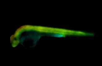
The method, termed as FLIM OPT, provides a powerful means to visualize cell signaling processes in their physiological context. Now, the performance of FLIM OPT was demonstrated in live transgenic zebrafish larvae using a genetically expressed FRET biosensor for Caspase 3. The researchers had generated a novel transgenic zebrafish line expressing a Caspase 3 FRET biosensor under the control of a ubiquitous promoter in a non-pigmented zebrafish. As soon as Caspase 3 is activated, it starts cleaving the biosensor, the fluorophores are separated, and the energy transfer between them decreases. Caspase 3 is a member of the cysteine dependent aspartate proteases, a family of proteases that cleave specific peptide sequences after an aspartate residue. It is a key enzyme in apoptosis, or programmed cell death, which is essential during development and homeostatic tissue turnover as well as during disease. If apoptosis becomes dysregulated it can lead to cancer, autoimmune disorders and neurodegenerative diseases.
Apoptosis was triggered in live zebrafish larvae by gamma irradiation at 24 hours post fertilization. Changes in Caspase 3 activation were monitored over time. The scientists observed significant apoptosis at 3.5 hours post irradiation, predominantly in the head region. Only 150 s were required to acquire the 3-D FLIM dataset, compared to 300 s required to image a single optical section using laser scanning confocal microscopy with time-correlated single photon counting. This should impose significantly less stress on the organism. The low light dose associated with wide-field imaging enables extended time-lapse studies to be undertaken with the potential to recover the zebrafish after imaging for further longitudinal studies.
The researchers believe that the presented approach could be useful in drug discovery, as zebrafish are an ideal model for drug screening. FLIM OPT could provide a way to visualize whole body responses to potential therapies in space and time. (Text contributed by K. Maedefessel-Herrmann)
 Cigarette compound exposure drives sorafenib resistance in hepatocellular carcinoma