May 12 2016
LaVision BioTec, developers of advanced microscopy solutions for the life sciences, report on the work of Ali Ertürk, a Group Leader at the Institute for Stroke and Dementia Research (part of the Klinikum der Universität München) at the Ludwig Maximilians University in Munich, Germany.

Dr Ali Ertürk, Group Leader of the Acute Brain Injury Research Group at the Klinikum der Universität München, part of the LMU.
Dr Ali Ertürk has been using light sheet and 2-photon microscopy in his research for a number of years during work in both the USA (with Genentech) and currently in Germany in the Klinikum der Universität München (KUM), of the Ludwig Maximilians University (LMU) in Munich. He is now Group Leader of the Acute Brain Injury Research Group where his main interest is in understanding key mechanisms leading to neurodegeneration after traumatic brain injury. At present, virtually nothing is known about how the initial trauma alters the brain structure over months/years and ultimately its function.
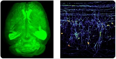
Left: Whole brain of a GFP-M mouse, which was cleared with 3DISCO method and then imaged with the LaVision UltraMicroscope.
Dr Ertürk describes the challenges he faces.
One of the main struggles in neuroscience in general is the difficulty to accurately analyze long connections in the brain using tissue sections which deliver only limited spatial information. We use a novel approach aiming at mapping the acute and chronic changes in the entire brain caused by small, well-defined brain lesions. To map the pathological brain, we utilize cutting-edge imaging techniques including high-resolution 3D imaging of the entire brain – that we recently developed – and in vivo 2-photon imaging. Subsequently, we screen for novel molecular players that are altered in chronically affected brain regions to halt secondary neurological problems.
Choosing the instrumentation from LaVision BioTec for his laboratory in Munich started with his experience gained using their first generation UltraMicroscope while in Genentech’s Department of Neuroscience. He was a member of the development team which discovered a highly reproducible and versatile clearing procedure called 3D imaging of solvent-cleared organs, or 3DISCO, which is applicable to diverse tissues including brain, spinal cord, immune organs and tumors. This has continued in Munich where he images entire transparent rodent brains. As Dr Ertürk says, the UltraMicroscope is the only commercial solution for this type of imaging where he looks at centimeter lengths of tissue. He also makes use of a two-photon microscope for higher resolution imaging of transparent organs albeit with a smaller field of view.
 Does motherhood influence brain aging? New research suggests a positive cognitive association