The major innovations in cancer diagnosis, therapy/palliative care and surgical intervention are too numerous to list here. But enormous strides have taken place in stem cell therapy, monoclonal antibody therapy and genetic screening.
In fact, over the last decade, such has been the impact of technology advancements, that a resultant increase in survival time is being seen in cancer.
Can you please give an overview of the patient pathway in cancer care?
If we were to start at the front end - of a patient admitted for surgery - he or she would be subjected to diagnostic imaging procedures. Generally, this would include CT/MRI/Ultrasound. More specific diagnostic imaging would either be by advanced optical diagnostics or advanced endoscopy. They would also receive blood diagnostics.
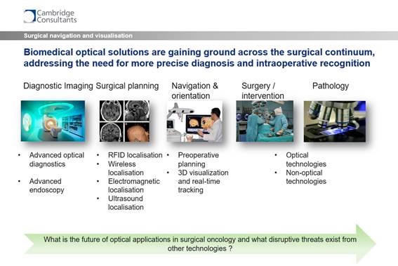
Those patients would then go through a process of surgical planning and procedures to localize the target tumor based on a number of technologies including RFID, wireless and electromagnetic localization and the use of ultrasound. Many software-based algorithms are used to build up a picture of that person's anatomy and the consequential cancer in that anatomy. Subsequently, there would be a surgical or intervention stage.
I'm just going to separate those two out. Surgery is quite clear. Using the prior imaging performed, the surgeon will remove the tissue from the suspected mass. Intervention is somewhat different. It's a less intensive procedure, involving specialized catheters delivered through a blood vessel leading to a tumor bed and an ablative procedure carried out to destroy the cancer. In principle this is very similar to a minimally invasive approach using laparoscopy or NOTES.
Surgical intervention will rely on optical technologies, to visualize local tissue changes, and non-optical technologies including X-ray, CT, and MRI type visualization to determine the overall anatomy of the tumor site.
Finally, a biopsy will be taken, which is sent to the pathology department, for specific histochemical analysis to determine the stage and type of cancer.
What advances have been achieved in surgical navigation and visualization for tumor detection?
I've given you a continuum there that starts with diagnostic imaging and goes to pathology via surgical planning, navigation and orientation, and surgery/intervention. The key areas that address the question you're asking involves navigation and orientation through to pathological assessment.
Cambridge Consultants has conducted a survey looking at the number of different research projects that are actively investigating surgical navigation and visualization programs for tumor detection. Worldwide, there are over seventy key groups that are investigating different technology applications.
The key advances can really be separated out into optical technologies and non-optical technologies. Several of these technologies have been developed from pre-existing diagnostic approaches. Rather than go into the nuances of that, actually let me go through the sorts of technology which are advancing our knowledge of surgical navigation and visualization in tumor detection and, more specifically, recognition of cancer invasiveness by evaluating the tumor margins.
For example, optical coherence tomography (OCT), which is a way of being able to provide 2D or 3D data, information or characterization. It is able to surface scan tissue to determine any changes in tissue morphology.
Although OCT has very high resolution, the downside is that it has a low level of penetration through tissues. Unsurprisingly, therefore, it has evolved from its use in various skin conditions, as it's a surface which is very accessible. Such skin diseases include basal cell carcinoma and melanoma.
Additionally, these technologies also have application in vascular imaging. In fact, OCT is a mainstream application in coronary artery imaging, by defining the extent of a lesion in an artery. Once again, the application uses superficial imaging within a vessel and does not require high penetration for the determination of lesion classification.
Similarly, a surface scanning application has been developed by NinePoint Medical (NVision VLE) for esophageal cancer obtaining FDA approvals in 2012. There is much attention on this technology to obtain more information on deeper lying pathologies, including different types of OCT and elastography including the use of needle probes to visualize deep lying pathologies.
In particular, the work from the OBEL laboratory in Australia (Prof David Sampson) has revealed stunning images of tumor margins. Similarly, the work of Prof. Stephen Boppart et al, from the University of Illinois has led to the development of a clinical spectral-domain OCT system for breast lumpectomy specimens in the OR. This author has also spun off a company called Diagnostic Photonics, which received FDA approval in 2014 for an interferometric synthetic aperture microscopy with OCT technology, called The Forsee imaging system.
These key developments are not just limited to OCT. Laser confocal endomicroscopy is proving to be a useful technology to track and diagnose tumours. Cellvizio® is a product developed by Mauna Kea for endoscopic imaging of cellular pathologies, and in the last year the company has received FDA approval for its use in surgical oncology.
Both NinePoint Medical and Mauna Kea have adopted the term optical biopsy, where cellular differentiation is being assessed without the need for removal of tissue, or as an alternative to frozen section at the point of tissue removal. For the moment, both optical biopsy and conventional biopsy are being conducted in parallel but assuming good sensitivity and specificity, it is likely that the need for complex tissue extraction prior to surgery will be superseded by these new technologies in the long term.
There are many other optical approaches providing visualization of tumors. For example, multi-spectral imaging, which requires more than one wavelength of light to differentiate the tissue types. There is some evidence, particularly from the University of North Carolina, that it can differentiate healthy and diseased pancreatic tissue.
Similarly, Raman spectroscopy, which is a spectroscopic method for looking at areas of cellular change in the body. At Cambridge Consultants, we have been involved in developing some Raman spectroscopy technologies for surgical oncology. This technology received high press coverage last year, when a Raman probe from Montreal was used as an imaging system in a patient undergoing surgery for brain cancer for the first time in the UK.
There is much current research activity with Fluorescence imaging, where injection of a fluorescence biomarker and use of an infrared light source is used to detect the localization of that biomarker in the body.
The disadvantage at the moment is that it's dependent on what we call fluorophores, which are fluorescent probes where you need a certain minimum of specificity and sensitivity for them to work. Most of the research is concentrated on developing highly specific cancer dependent fluorophores. The amount of effort required is such that the cost of developing such a diagnostic imaging agent would be somewhere around a hundred million dollars.
The return on that investment depends on the target tissue and the larger target patient populations. However, I have no doubts that in twenty years, it will be a mainstream application for cancer detection during surgery.
It's all very well finding the tumor during surgery, but actually the most important aspect of any diagnostic during surgery is not just localizing the tumor, but making sure that the margin, which is the area between the tumor and normal tissue, is free of any cancerous cells. One cell left behind will, in no time at all, exponentially grow. Therefore, it's key for the surgeon to recognize that the bed that's been left after the tumor has been removed is free of cancer cells.
With many of these technologies such as OCT, multi-spectral imaging and Raman spectroscopy, the fundamental focus is to make sure that there are clear tumor-free margins in those patients from whom tumors have been taken.
In what ways has the use of imaging for guidance during surgical interventions been limited so far?
The first part of that is quite straightforward because most soft tissue imaging that is capable of being performed is performed by MRI. However, this technology, in the main, is just too bulky to use during a routine surgery. There are some systems out there now which are being developed for use in the operating room, but at the moment, they are primarily diagnostic in nature.
Currently MRI or CT images are taken pre-surgically and interventions recorded as an overlay of the pre-surgical images.
Until now, there just haven't been the tools available to determine whether a surgeon has removed all cancerous tissue at the time of the surgery. The way it's done currently is that the tumor is removed sent to a pathologist who evaluates the margins. A cold section is usually taken at the time of surgery, which a pathologist processes and reports back to the surgeon. However, this approach is actually still fairly crude. The answers are still unclear until a full histopathology is conducted, which may take several days.
Naturally, the patient's already been treated or undergone surgery and often this results in the patient having to go back for repeat surgery.
Can you please give an overview of the iKnife? How does this ‘intelligent’ knife work and what information can it provide the surgeon with?
The iKnife was developed by a group at Imperial College. It is an intra-operative tissue identification technology using a technique known as Rapid Evaporative Ionisation Mass Spectrometry (REIMS).
The innovative iKnife hand-held sampling device produces information-rich vapor directly from the surface of the sample, which when analyzed by a time of flight (Tof) MS, provides analysts with an accurate molecular profile in seconds.
Cancer tissues have different molecular properties than ordinary tissues and so it's a very simple way of being able to determine, with each movement of the iKnife, exactly what molecules exist.
In studies where they’ve used this, when trying to profile cancerous and normal tissue, the authors have quoted that they've had a 100% success in ninety-one patients.
At what stage of development is the iKnife?
This technology was acquired a couple of years ago by a company called the Waters Corporation, who are now providing marketing materials on the product.
How personalized do you think surgery could become with technologies such as the iKnife?
I don't think that the iKnife or any of the other technologies discussed aids in the personalized nature of surgery. I think the personalized nature comes in when certain diagnostic tests are applied to cancer patients to assess what types of therapy those patients should have.
There are lots of diagnostic tests out there that look at circulating tumor cells or free DNA measurements which could define the patient upfront and therefore allow for pharmaceutical treatment of that patient.
You can then select the patient for whom one therapy would be more beneficial compared to another. I think that level of personalization is very exciting, but actually removed a bit from the levels of surgical interventions.
What do you think the future holds for emerging technologies in cancer care?
We must think about what non-optical technologies are out there and there are two examples.
The iKnife is one of them. It's not an optical system; it's a spectroscopic system. The other one is a product called MarginProbe. This is being developed by Dune Medical in the US and is a way of being able to define cancer or normal tissues by means of an electromagnetic measurement called fringe field sensing. Essentially, it is a probe that looks at the impedance changes between cancerous tissue and normal tissue.
It has been used in breast cancer patients and has been shown to reduce the re-operation rate. That is when a lump is taken from a patient using the MarginProbe to determine the level of cancer in the tumor margins. Re-operation rates for those patients have been shown to be reduced by 56%, which is a huge saving to the surgical world.
Coming back to your question about what the future is for these technologies, all are trying to address the same thing, which is to remove cancer cells from the margin of tumor beds in patients, allowing performance of the diagnosis and surgery at the same time, reducing delays in treatment and obviating the need for repeat surgery.
I've said about MarginProbe reducing the re-operation rates by 56%, which is a fantastic outcome, but actually it's not the real outcome. The real outcome has to be survival times. As some of these technologies have really only received their regulatory clearance in the last couple of years, it's going to be some time before we understand whether any of them enhance the survival times of cancer patients for any given cancer.
Where can readers find more information?
About Tim Clay
 Tim Clay is an Associate Director at Cambridge Consultants specializing in medtech product strategy and development.
He holds an M.I.in Biology and an M.Phil in Physiology from the University of London.
At Cambridge Consultants, Tim oversees USP product development and has responsibility for portfolio enhancement including M&A and technology acquisition as well as product launch and growth strategies.
 Oxybutynin shows promise in managing hot flashes for prostate cancer patients