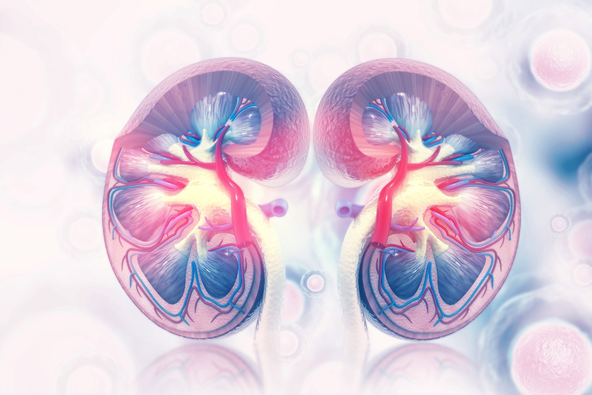
About 50% of the adult population throughout the world over the age of 70 years has chronic kidney disease (CKD) and renal fibrosis. Both CKD and renal fibrosis begin as pyroptosis, during which a certain type of regulated cell death (RCD) occurs in response to inflammation to restore normal function to the organism ultimately.
 Study: Pyroptosis in renal inflammation and fibrosis: current knowledge and clinical significance. Image Credit: crystal light / Shutterstock.com
Introduction
Renal fibrosis is the end stage of any chronic inflammatory disease of the kidneys.
CKD passes through three stages in its terminal phase, including wound healing, inflammatory, and proliferative phases, during which tissue remodeling occurs, with loss of normal renal anatomy and function. While epithelial and endothelial cells are involved in triggering inflammation during the second phase by recruiting macrophages and other inflammatory cells, macrophages also promote fibrosis in the last phase. This switch in function is mediated by a corresponding change in the cytokines released by the macrophages.
Fibroblasts convert into myofibroblasts, with both types of cells proliferating during chronic inflammation. These cells produce multiple inflammatory chemokines that cause fibrotic changes. Simultaneously, the extracellular matrix accumulates to lead to interstitial fibrosis of the kidney ultimately.
The role of pyroptosis
Various pattern-recognition-receptors (PRRs) exist on epithelial and innate immune cells that are activated when their encoded germline recognizes a specific microbial pattern. Different types of PRRs recognize damage-associated molecular patterns (DAMPs), pathogen-associated molecular patterns (PAMPs), or both and initiate various types of responses, including inflammation.
The goal of pyroptosis is to inhibit infectious pathogens through a potent inflammatory response. Pyroptosis produces significant swelling of affected cells, which break open and cause inflammation.
Cell death occurs during pyroptosis due to the activation of inflammasomes that cause gasdermins (GSDM) to break apart. Subsequently, holes develop within the membrane, which allows the pro-inflammatory signaling molecule interleukin (IL)-1β/18 to escape. Two pathways have been observed in pyroptosis, which includes classical and non-classical pyroptosis that are mediated by caspases-1 and 4/5/11, respectively.
What do GSDMs do?
GSDMs are associated with different disorders, such as autoimmune disease with GSDMA. GSDMD was the first molecule to be linked to pyroptosis and appears to be the main effector molecule.
The NLRP3 inflammasome is a molecular platform that is closely linked to the start of pyroptosis. This molecule responds to multiple stimuli by becoming activated.
NLRP3 leads to a protective inflammatory response when activation occurs following infection in an effort to support the survival of the organism with minimal damage. However, NLRP3 may also cause damage when excessively activated.
Several steps are involved in activating the NLRP3 inflammasome, including its priming by microbial toll-like receptor (TLR) ligands and subsequent inflammasome complex assembly. This leads to the cleavage-activation and secretion of IL-1β/18.
In the classical pathway, the NLRP3 inflammasome is activated through TLR-PAMP binding, thereby promoting NLRP3 gene expression.
This is followed by inflammasome assembly through ATP binding by causing membrane porosity, the formation of crystals such as uric acid or silica causing lysosomal rupture with the release of cathepsins, or mitochondrial DNA with other molecules triggering reactive oxygen species (ROS).
In the non-classical pathway, lipopolysaccharides (LPS) enter the cell and directly activate caspase enzymes, thus activating the inflammasome and causing GSDMD cleavage.
These findings indicate that GSDM inhibitors could treat these diseases by preventing pyroptosis. However, additional research is needed to elucidate the function of pyroptosis and GSDMs in triggering renal inflammation and fibrosis.
Therapies countering pyroptosis
Various strategies have been considered to mitigate pyroptosis and prevent fibrosis. These include inhibitors of inflammasome assembly, caspase-1 activation, inflammatory cytokine release, GSDMD activation, and earlier pathways.
Among the therapeutics that have been studied include MCC950, which prevents NLRP3 activation, tranilast, which stabilizes the cell membrane, BAY-11-7082, an NLRP3 ATPase inhibitor, fucoidan, which blocks NLRP3-linked pyroptosis, and phloretin, which inhibits NLRP3 inflammasome activation.
Caspase-1 activation is blocked by Z-YVAD-FMK and VX-765. IL-1β inhibitors include anakinra and canakinumab.
Disulfide directly inhibits human GSDMD to inhibit the inflammatory signaling pathway ultimately. The inhibition of inflammasome activation caused by ROS is another potential target, such as by NADPH oxidase inhibitor complanatoside A.
What are the implications?
While researchers have acquired important information on how the inflammasome is activated, its significance during renal fibrosis, and the critical role of GSDMD, many additional studies are needed to understand pyroptosis and its impact on renal inflammation and fibrosis.
We believe that pyroptosis-focused research will open up new perspectives for diagnosing and managing renal fibrosis.”