These findings represent the first description of a human coronavirus exposing its S1B domains on demand, thereby suggesting that the mode of beta coronavirus attachment to host cells is even more complex and sophisticated than previously appreciated.
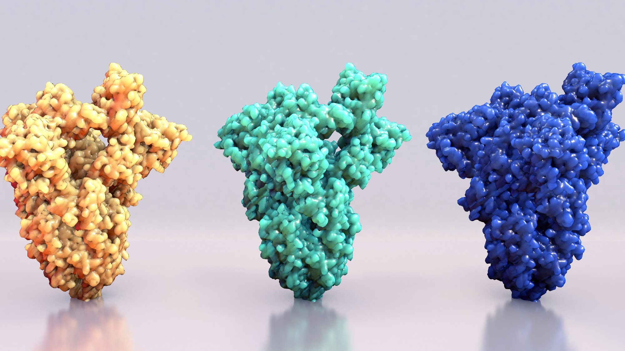 Study: Sialoglycan binding triggers spike opening in a human coronavirus. Image Credit: Design_cells / Shutterstock.com
Embecovirus and the importance of CoV spike proteins
Prior to the emergence of the severe acute respiratory syndrome coronavirus 2 (SARS-CoV-2), four main coronaviruses (CoVs) species colonized humans globally. Two of these viruses, namely HKU1 and OC43, crossed over to humans from rodent reservoirs and are unique from other CoV species, as they require cell surface glycans as primary receptors. Both HKU1 and OC43 belong to the betacoronavirus subgenus Embecovirus.
Spike proteins, also known as peplomer proteins, form rod- or club-shaped projections from the surface of an enveloped virus. Spike proteins are characterized as having typically large external ectodomains, a single transmembrane domain that anchors the protein in the viral envelope, and a short tail in the interior of the virion.
Spike proteins play an essential role in viral attachment and subsequent entry into host cells. Most spike proteins have a binding site for cell-surface receptors, which is usually located at the tip of the spike, that recognizes host cell surface receptors and preferentially binds to them for cell entry.
Embecovirus spike proteins bind to 9-O-acetylated sialosides (9-O-Ac-sialosides). This binding is highlighted by members of the subgenus that code for haemagglutinin esterase, an additional envelope protein not found in other CoV strains. Haemagglutinin esterase is a type of sialate-O-acetylesterase that functions as a receptor-destroying enzyme.
CoV spike proteins belong to a subclass of proteins known as homotrimeric class I fusion proteins, which can be divided into amino- (designated ‘S1’) and carboxy-terminal (designated ‘S2’) regions. S1 protein domains are involved in mediating receptor binding, whereas S2 domains comprise the fusion machinery of the CoV spike protein.
In Embecoviruses, attachment to the 9-O-Ac-sialosides occurs through a well-conserved host receptor binding site found in the S1A domain. Recent studies have found evidence for a second domain, S1B, that may serve as a secondary receptor, as demonstrated by the mapping of virus-neutralizing antibodies map to subdomain S1B2.
The spike proteins of SARS-CoV, SARS-CoV-2 and Middle East respiratory syndrome coronavirus (MERS-CoV) occur in different conformations with their receptor-binding S1B domains either partially buried between neighbouring protomers (‘closed’ or ‘down’) or with one or more S1B domains exposed (1-, 2- and 3-up, ‘open’)”
Since spike and other cell-surface virus proteins are used by the host immune system to identify and irradicate infections, the transition between open and closed S1B states allows the virus to safeguard itself against the host immune response when closed while retaining its infective potential in an open state. Barring rare exceptions, the pre-fusion spike proteins of all CoVs have been exclusively observed in the closed state.
Given that both open and closed states are known to science, research into the triggers/specific cues of transition between S1B states would shed light on the mechanisms by which CoV strains escape host immune detection and achieve the high virulence they are observed to.
About the study
In the present study, researchers investigate the cues modulating the shift between open and closed states in human HKU1 CoV. To this end, cryo-EM was used to evaluate the structure of HKU1 spike proteins and the relevant protein domains.
The researchers began by expressing and purifying trimeric HKU1 spike ectodomains by expressing a sequence of the HKU1-A spike protein in a pCG2 expression vector. This construct was subsequently incorporated into HEK293T transient vector cells, whereas the spike glycoprotein was purified using affinity chromatography.
The resultant purified spike proteins were prepared for cryo-EM using QuantiFoil R1.2/1.3 grids, which were blotted and plunge-frozen in liquid ethane. A total of 4,207 videos (apo spike protein) and 4,065 (holo spike protein) were generated at a resolution of 0.415 Å per pixel using a cyro-transition electron microscope (cryo-TEM).
Single-image particle processing involved patch motion correction processing, during which micrographs with a contrast transfer function (CTF) resolutions of < 10 Å were excluded from further analyses.
A template HKU1-A spike protein homology model was created referencing the protein structure of a previously described Embecovirus OC43 spike structure. Due to differences between OC43 and HKU1-A S1B crystal structures, the template model was improved by substituting the OC43-equivalent S1B structure with a HKU1-A S1B crystal structure derived from cryo-TEM data analyses. N-linked glycans were subsequently constructed using the Coot carbohydrate model, following which manual inspections and optimizations were performed.
Molecular dynamics simulations were devised using data obtained from cryo-EM structure images. N-glycans were then attached to the protein using quantitative data from site-specific N-linked analysis of the analyzed HKU1 spike protein.
Visualization of the open and closed states of the HKU1 protein was achieved using the UCSF ChimeraX software, with spike protein coloring carried out in Consurf. PDBePISA was used to calculate spike interface areas, and the UCSF Chimera MatchMaker tool was used for obtaining root mean square deviation values.
Study findings
The present study addressed the confound of no prior open-state CoV spike protein observations by using cryo-TEM and molecular dynamics modeling to describe that a specific cue of binding of the disialoside-based receptor 9-O-Ac-Sia(α2,8)Sia to S1A can result in the non-spontaneous opening of the hitherto close spike protein configuration. Since this transition from the closed to open state exposes S1B, thereby allowing it to bind to proteinaceous cell surface receptors, facilitating infection, this result presents an important step for elucidating the mechanisms that allow CoV viruses to escape host immune reactions while retaining their infective potential.
TEM and modeling data revealed four distinct protein structures alongside the transition from fully closed (apo) to fully open (holo) spike protein conformation. In the initial transition step, S1A disialoside binding converts the apo state into a still closed yet primed for the S1B transition state. Despite being extremely transient and short in duration, rapid high-resolution images were able to confirm this as a distinct state not previously described.
Our findings suggest a causal mechanistic relationship between the disialoside-induced conformational changes in e1, S1A1 rotation, the remodelling of the S1A–S1B interface and S1B expulsion. Yet, we note that the topology of the e1 element in our HKU1-A spike apo structure is atypical and differs from that in the spike protein of HKU1-B and those of betacoronavirus-1 variants OC43, bovine CoV and porcine haemagglutinating encephalomyelitis virus”
These results suggest that the mode of CoV attachment to human cells may be far more sophisticated and complex than previously assumed. The study findings also imply the presence of dual receptor usage and S1A priming as a modality to escape host immune detection.
Journal reference:
- Pronker, M. F., Creutznacher, R., Drulyte, I., et al. (2023). Sialoglycan binding triggers spike opening in a human coronavirus. Nature; 1-6. doi:10.1038/s41586-023-06599-z
 Long COVID symptoms change for 15 months after infection