Introduction
Protein aggregation refers to the clumping together or accumulation of mis-folded proteins. This aggregation can occur during the production, administration, storage, and shipping of therapeutic protein compounds, which can compromise their stability, efficacy, and safety. The size of protein aggregates can range from a few nanometers to hundreds of micrometers. The size and abundance of any protein aggregates must be determined during each step of the production of therapeutic proteins. To this end, a number of light scattering techniques have been developed to characterize protein aggregates, enabling their composition in therapeutic formulations to be determined.
Dynamic and Static Light Scattering Techniques
Dynamic light scattering (DLS) and static light scattering (SLS) are techniques that have been applied in the characterization of protein aggregates ranging from several nanometers (monomer and low-order oligomer) to micrometers (high-order aggregate and aggregate particles) in diameter. The strength of the light scattering signal is relative to both the concentration and molar mass of the species in solution.
Light scattering detectors are therefore extremely sensitive in the detection of large aggregates, even at small quantities. In addition, contemporary DLS and SLS detectors can be combined with on-line fractionation techniques such as field-flow fractionation (FFF) and size exclusion chromatography (SEC) to allow superior resolution and excellent characterization of the various sub-species present in protein formulations.
Although both SEC and FFF separate molecules based on their hydrodynamic volume, SEC is generally used to separate proteins, whereas FFF can separate both proteins and nanoparticles. In most laboratories, SEC is used to isolate and measure the stable protein isoforms from aggregates and fragments.
Static light scattering can be right angle, low angle, or multi-angle LS (MALS). Of these, MALS is the most precise, flexible and widely used method for comprehensive characterization of polymers and biopolymers (Figure 1).
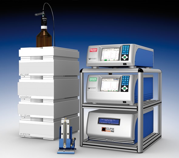
Figure 1. Separation of protein monomers and aggregates is accomplished by SEC or asymmetric flow field-flow fractionation (AF4). On-line analysis of molar mass and size uses MALS, DLS, UV/Vis absorption (UV), and differential refractometry (dRI).
MALS detectors may be used either in batch mode or together with FFF or SEC separation. Importantly, the addition of a MALS detector to FFF or SEC gives the absolute molecular weight (i.e. independently of density and geometry) and SEC or asymmetric flow field-flow fractionation (AF4) is used to separate protein monomers and aggregates.
On-line analysis of size and molar mass uses DLS, MALS, differential refractometry (dRI), and UV/Vis absorption (UV), as well as information such as number density data for aggregate particles, root mean square radius (Rg, when >10 nm), or stoichiometry of protein conjugates, among other parameters.
Characterization of Aggregate Particle Structure
In order to determine the structure of aggregate particles or measure protein radius less than 10 nm, DLS can be combined with MALS, downstream of the FFF or SEC, to give the hydrodynamic radii (Rh) of the eluting species.
In the case of proteins, combined information from DLS (Rh) and MALS (molecular weight) can be valuable in understanding molecular conformation and folding states. In the case of larger species, the ratio of the hydrodynamic radius and root mean square radius, from DLS and MALS, respectively, can not only be used to determine particle shape, but to measure mass distributions within the particle.
In order to determine the distribution of protein aggregates, DLS or MALS measurement is usually carried out in batch mode without separation. The main application mode of DLS is batch analysis, which monitors aggregation as a function of several different parameters, such as temperature, concentration, time and buffer composition. Unlike MALS, the concentration does not need to be known for DLS. These factors, combined with the ability to make rapid measurements, make batch DLS a valuable tool for high-throughput screening of formulation stability, particularly when using automated DLS instruments with well plates.
Although batch DLS is extremely sensitive to the presence of trace amounts of aggregate, the resolution of sub-species using this technique requires that their radii vary by a minimum factor of four. The development of low-order oligomers can only be monitored through an alternative metric as a proxy. However, this is not a limitation with on-line DLS after fractionation.
As with DLS, batch MALS can be used to measure average molecular weight with minimal resolution of different species in a sample. However, MALS is much more sensitive than DLS when evaluating lot-to-lot variability. Under favorable conditions, batch MALS can easily detect a 1% change in the average molecular weight caused by a slight variation in either the amount or type of aggregates in solution. When calculated at different concentrations, data from batch MALS can be used to identify protein self-association as well as to resolve and measure different types of oligomers present in a sample across a wide concentration range.
Conclusion
Static and dynamic light scattering detection techniques are often used to characterize molecular physical properties during the development of therapeutic formulations. Both techniques can be employed for either batch analysis or for enhanced detection and characterization together with different fractionation methods.
Light scattering following fractionation allows resolution and characterization of different aggregates in a protein solution that are permanently associated. However, occasionally, it is the separation process itself that changes the sample’s aggregation state or content. In contrast, batch analyses allows high-throughput screening for solution conditions that encourage protein aggregation and for characterizing different specific and non-specific associations. These complimentary techniques each provide their own valuable information, which combine to offer a reliable means of fully characterizing aggregation phenomena in protein-based formulations.
Reference
Reprinted with permission from "Methodologies for characterising protein aggregation" by Michelle Chen, 2013. Sp2 InterActive, November/December 2013, pg. 20-22.
About Wyatt
 With a long history of excellence in scientific instrumentation, Wyatt Technology is the recognized leader in innovative light scattering instruments, accessories, software and services for determining the properties of macromolecules and nanoparticles in solution. Wyatt provides cutting-edge solutions for in-line multi-angle static light scattering (SEC-MALS), field-flow fractionation (FFF-MALS), composition gradients (CG-MALS), high-throughput and traditional dynamic light scattering (DLS), electrophoretic mobility via phase-analysis light scattering (MP-PALS), differential refractometry and differential viscosity. With a staff composed of 20% Ph.D. scientists and many more dedicated and experienced support personnel, Wyatt's aim is to delight the customer with the best products, training, customer support and service available in the industry.
Sponsored Content Policy: News-Medical.net publishes articles and related content that may be derived from sources where we have existing commercial relationships, provided such content adds value to the core editorial ethos of News-Medical.Net which is to educate and inform site visitors interested in medical research, science, medical devices and treatments.