In a recent study posted to the bioRxiv* preprint server, researchers explored the association between severe acute respiratory syndrome coronavirus 2 (SARS-CoV-2) spike protein and burst activities in neurons.

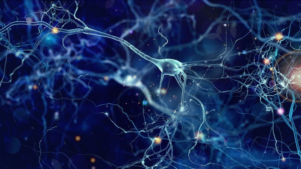
Study: SARS-CoV-2 Spike Protein Reduces Burst Activities in Neurons Measured by Micro-Electrode Arrays. Image Credit: whitehoune/Shutterstock.com

 *Important notice: bioRxiv publishes preliminary scientific reports that are not peer-reviewed and, therefore, should not be regarded as conclusive, guide clinical practice/health-related behavior, or treated as established information.
Background
The primary components of the SARS-CoV-2 structure are envelope (E), spike (S), membrane (M), and nucleocapsid (N) proteins. This research focuses on the impacts of the S protein. The S protein facilitates virus attachment and entrance into the host cell. The S1 subunit interacts with the angiotensin-converting enzyme 2 (ACE2) receptor present in the intestinal and lung cells.
Within the brain, ACE2 is predominantly expressed in the brain stem and regions whose primary function is to regulate blood pressure and cardiovascular function. While neurological signs have been documented in some, not all, coronavirus disease 2019 (COVID-19) patients, the precise mechanism by which viruses affect neuronal cells is still unknown and, thus, a subject of investigation.
About the study
In the present study, researchers quantified the neurological phenotypes induced in neurons by the SARS-CoV-2 S protein.
In this study, the S1 and S2 subunits of the spike protein were evaluated separately to determine if they elicited any neurological phenotypes as estimated by the micro-electrode arrays (MEAs). On day zero, neurons obtained from newborn P1 mice were treated with recombinant SARS-CoV-2 S protein and S1 and S2 S2 subunits. The team assessed the data using an algorithm devised in-house. Characteristics like the number of bursts per electrode, their duration, frequency, and the number of spikes per burst according to the treatment condition were also quantified.
The team also determined whether the S1 subunit influences mature neurons during cell exposure. The neurons were treated with similar S1 concentrations on day 12. This was followed by exposure to the same S1 concentration for seven consecutive days. The team then performed a rescue experiment to ascertain if this neuronal phenotype is reversible. Before administering S1 to neurons on day zero, a human monoclonal anti-S1 antibody was sampled and neutralized using the antibody. To test the hypothesis that the S1 receptor-binding domain (RBD) may be the reason for burst reduction, the team collected and assessed purified recombinant RBD.
Results
The study identified the number of pulses per electrode as the most prominent characteristic that differentiated the spike protein-treated wells from the control wells. The S1 subunit substantially lowered the number of bursts per electrode, whereas the S2 subunit did not exhibit the same degree of reduction. The study suggested that S1 is responsible for decreasing burst activities of neuronal populations when cells are exposed early in the course of development. However, there was no discernible difference in burst activity between S1-treated and the control wells. Therefore, the data indicated that the S1 subunit affected neurons only when the cells were exposed during the earliest stages of development.
S1 neutralized by antibodies did not result in a significant decrease in burst activity compared to the control, whereas the conventional S1 treatment on day zero did reduce burst activity. The data as well as the p values suggested that the anti-S1 antibody reversed the impact of S1 on bursting activities. Overall, the rescue experiment provided compelling evidence that S1 was able to suppress burst activities when exposed to cells early in their developmental course. Comparable to the S1 data, the team identified a significant reduction in surge activities. The outcome strongly suggests that the RBD itself is sufficient to suppress surge activities.
Conclusion
The study findings demonstrated a causal relationship between the SARS-CoV-2 S1 protein and in-vitro burst trends in neuronal populations, which can be reversed by antibody treatment. The study also noted that the RBD may be accountable for the suppression of neuronal signals. This study also indicated that neutralizing S1 restores neuronal discharge activities to control levels. Furthermore, the antibody rescue experiment confirmed the role of S1 in suppressing burst activities and highlighted the protective function of anti-S1 antibodies as well as the significance of RBD in modulating neuronal phenotypes.
The researchers believe that the study findings provide new insight into the activity of SARS-CoV-2 S proteins beyond their well-established functions in viral attachment and entry. This discovery may shed light on crucial aspects of SARS-CoV-2 infection, patient care methods, and future vaccine and antiviral development.

 *Important notice: bioRxiv publishes preliminary scientific reports that are not peer-reviewed and, therefore, should not be regarded as conclusive, guide clinical practice/health-related behavior, or treated as established information.