Their results indicate that exposure to depression may be associated with functional changes, with variation seen based on which definition of depression is used.
 Study: Lifetime Exposure to Depression and Neuroimaging Measures of Brain Structure and Function
Background
Depression or major depressive disorder (MDD), which is associated with feelings of excessive guilt, loss of interest, and low mood and may affect more than 10% of people during their lifetimes, can severely affect one's quality of life while increasing one's risk of self-harm and suicide.
Efforts to understand the neurobiological underpinnings of depression can improve treatment for people experiencing this challenging issue.
Previous studies have found that MDD can reduce hippocampal volume and increase amygdala activity, particularly during emotional tasks. However, meta-analyses indicate a lack of convergence across studies and small effect sizes which could be due to the variability in what definition of depression is applied.
It seems likely that some brain alterations may be either a predisposing factor for MDD or a consequence of its diagnosis and treatment, but further investigation is needed to understand whether these are persistent and whether depression can affect the function and structure of the brain in one's later life.
About the study
In this study, researchers used six different criteria that are used to define lifetime exposure to depression, namely self-reported depression, help-seeking for depression, antidepressant use, two definitions from clinical literature, and one based on a diagnostic interview score.
To be included in the study, individuals had to meet at least one of these six criteria and were then stratified into six groups depending on how many criteria they met. This ensured that each participant was part of only one group and that there were no overlaps.
These groups were then matched to control or comparison groups of 'healthy' individuals who did not have a history of mental illness, psychosis, behavior disorder, or any other nervous system condition. Demographic variables were included as covariates.
The imaging data used in the study were resting-state functional magnetic resonance imaging (MRI) and T1-weight structural images. These were pre-processed before analysis. Relative gray matter volume (GMV) and functional indices such as the fractional amplitude of low-frequency fluctuation (fALFF), local correlation (LCOR), and global correlation (GCOR) were calculated.
The groups of individuals with depression were compared to the healthy control groups using two-sample, two-tailed t-tests, and effect size computation.
Findings
The study included 20,484 people who met one or more criteria of depression exposure in their lifetime and 25,462 health controls. People with a history of depression were predominantly female (61.7%), 64 years old on average, and had an average of 16.7 years of education. People in the control group were less likely to be female (44.7%), were 65 years old on average, and had 17 years of education (mean).
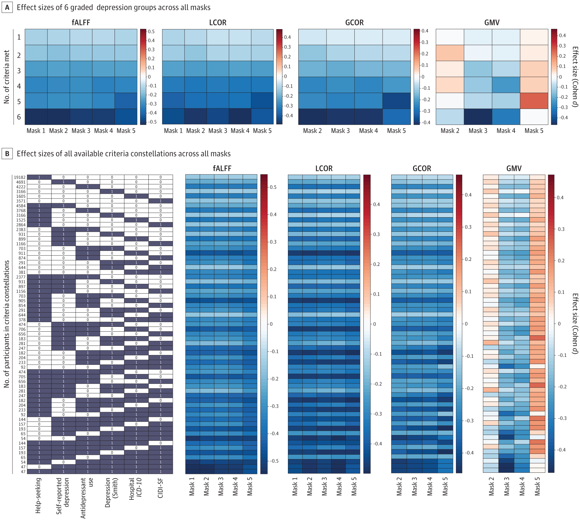 A, Masks indicate the number of criteria met (eg, mask 1 indicates meeting 1 criterion; mask 5, meeting 5 criteria). Effect sizes were calculated in all depression stratum–mask pairs. B, The dark element indicates that the column contains corresponding criteria. Other blocks show the effect size values (Cohen d). Red indicates increased imaging measures in depression group, and blue indicates reduced imaging measures in depression group. CIDI-SF indicates Composite International Diagnostic Interview Short Form; fALFF, fractional amplitude of low-frequency fluctuations; GCOR, global correlation; GMV, gray matter volume; ICD-10, International Statistical Classification of Diseases and Related Health Problems, Tenth Revision; LCOR, local correlation.
Compared to the health control group, there were significant differences in structural and functional measures for people with depression, including consistent reductions in GCOR, LCOR, and fALFF. However, there was no significant difference in GMV. Functional alterations were noted in the cerebellum, prefrontal cortex, middle temporal cortex, parietal cortex, occipital cortex, and fusiform gyrus.
LCOR and fALFF alterations were noted in the bilateral postcentral and precentral gyrus, while reductions in GCOR were observed in the insula, pre-cuneus, lingual cortex, superior temporal cortex, and middle inferior temporal cortex. Effect sizes were largely consistent with these findings.
Conclusions
Researchers systematically assessed the associations between brain functional and structural measures and lifetime depression exposure. A broad observation was that the application of different depression definitions led to differences in structural and functional alterations; however, applying more restrictive definitions led to higher functional but not structural impacts.
An important consideration is that, among the criteria, antidepressant use is highly correlated with a diagnosis of depression, limiting the causal interpretations of the alterations that have been observed. However, depression is treated not only with medication but also with psychological therapies such as stimulation and cognitive behavioral approaches.
These results demonstrate how observed imaging outcomes are affected by which definition of depression is applied. Researchers may use less strict definitions so as not to decrease sample size and lose statistical power, but this may lead to dilution in the observed imaging alterations associated with depression.
Further explorations should investigate how more stringent criteria can be applied with a focus on which definitions of depression would be most beneficial for improving depression treatment and outcomes.
Journal reference:
- Lifetime exposure to depression and neuroimaging measures of brain structure and function. Wang, X., Hoffstaedter, F., Kasper, J., Eickhoff, S.B., Patil, K.R., Dukart, J. JAMA Network Open (2024). doi:10.1001/jamanetworkopen.2023.56787, https://jamanetwork.com/journals/jamanetworkopen/fullarticle/2815247
 Small molecule targets glioblastoma oncogene in preclinical studies