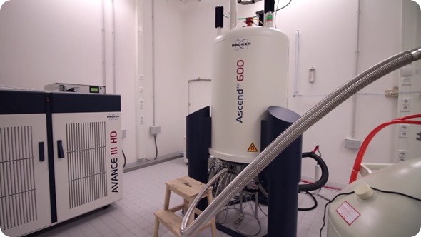
Nuclear magnetic resonance (NMR) spectroscopy is a powerful analytical tool widely used in chemical research to investigate the structure and dynamics of molecules.
At the Leibniz University of Hannover, a team of researchers have been using a multidisciplinary approach that combines NMR, biochemical, biophysical and computer-based methods to investigate the structure of high-molecular weight complexes that cannot be tackled using NMR alone.
Protein Complexes with NMR
The group is concerned with two main lines of research. One is the structure and function of ribonucleoprotein complexes (RNPs) that have catalytic activity in RNA metabolism and gene expression regulation. The other is the interaction between small molecules and their large, macromolecular receptors, for the development of methodologies for structure-based drug design.
In an interview with News Medical, team leader Prof. Dr. Teresa Carlomagno described a complex the group have recently investigated that methylates ribosomal RNA (rRNA). She said rRNA and, in fact, RNA in general, undergoes modification at different positions: “One of the most widespread of these chemical modifications is methylation at the 2’-O position of the ribose and the cell goes through a lot of trouble to achieve this modification.”
The cell has complicated machinery in place that involves guide RNAs recognizing rRNA sequences that require methylation. Each of these guide RNAs recognize two rRNA sites. The researchers presumed that if this was functionally significant and the two methylated sites control each other, the structure at the two methylation sites would need to differ.
However, one structure published by other researchers and established by X-ray crystallography showed that the two rRNA binding sites were in exactly the same environment and had the same structure.
Further investigation by Carlomagno’s group, using a combination of NMR and small-angle neutron scattering, revealed a structure that differed to the published one. The researchers had shown, for the first time, that the rRNA recognition sites in fact exist in two different environments, suggesting that enzymatic methylation of the sites occurs in sequence, rather than simultaneously.
The team performed functional assays in the NMR tube that showed the two RNA sequences were indeed methylated depending on each other, with the “leader” sequence requiring methylation in order for the other to be methylated and released. This meant sequential regulation was occurring and, next, the researchers have been designing cell biology experiments to verify the physiological meaning of this control.
We think this is a process the cell uses to efficiently and quickly allow for ribosome folding. In fact, if you eliminate methylation from the cells, they die.”
Prof. Dr. Teresa Carlomagno
She also pointed out that the functional experiments could only be designed as a result of the information gleaned from the discovery of their structure and the sequential methylation: “This is something that neither the X-ray structure nor the electron microscopy structure were able to reveal. The other techniques only take a static picture of the complex, whereas we can follow the whole cycle.”
Carlomagno went on to describe a methodology the team have recently developed to support structure-based drug design. The group had been approached by pharmaceutical companies wanting to know how certain drug leads bind to receptors. In order to do this, the researchers needed the drug to crystallize with the receptor, but with large receptor molecules, this is not always possible.
The team decided to look at the ligand only. This ligand retains a “memory” of the receptor-bound state, once in the free state. This free state can tell the researchers the conformation the ligand adopts when it binds to a receptor. However, it cannot tell you how the binding occurs.
This led Carlomagno’s group to develop a new NMR-based methodology called InPharma. InPharma employs two ligands that bind to the receptor in an alternating manner, continuously exchanging their position in the binding pocket. Information left in the pocket by the first ligand is then “learned” by the second ligand when it binds.
“By making the two ligands talk in this way, you know which ligand atom had been close to which part of the receptor by reading it out in the parts of the ligands that have been able to communicate among each other,” explained Carlomagno. This information is extracted using bioinformatics and then used to find out which parts of the receptor the ligands have seen and how they communicated with the receptor. “By developing this methodology, we have found a way to get the binding modes of these ligands to receptors in cases where a crystal structure cannot be solved.”
Carlomagno added that she is very happy with the product and pharmaceutical companies have already been reporting on InPharma data that they have managed to acquire without the help or advice of the group.
 Collaborative approaches to honey adulteration detection