Fluorescence imaging is a quantitative and sensitive technique which has become integral to clinical applications and life sciences research. Visualization of bacterial colonies grown on agar plates is one form of fluorescence imaging.
This method can be applied to a number of investigations, including studying protein-protein interactions, screening for gene insertions or mutations, or verifying fluorescent protein expression, this is in addition to the detection and evaluation of microbes in water, soil, and food1,2.
With current innovations in analysis technology and colony imaging, utilizing vectors containing fluorescent reporter genes ligated with a gene of interest provides a more robust and convenient technique to count and select transformed cells or screen for mutations.
Liquid microbe cultures that contain these vectors can be diluted and plated on agar plates. The plated microbes form colonies after incubating at the appropriate temperature and time depending on the microorganism used, each growing from one progenitor cell.
Next, colonies are counted to establish the number of colony-forming units (CFU) and the original concentration calculated. Conventional techniques rely on manual counting, usually employing grids that draw on an agar plate to ensure colonies are counted accurately.
Fluorescence imaging provides a less time-consuming, more accurate way to assess colony growth. Compact, stand-alone imaging systems such as the UVP GelSolo supply high contrast image acquisition to ensure simple and effective identification of labeled colonies.
The imager streamlines fluorescence imaging with a touch screen with intuitive VisionWorks image acquisition and analysis software and live view capabilities. This article outlines the colony counting process with specific emphasis on imaging of plates containing fluorescent colonies.
Simulation of fluorescence imaging will be done by utilizing a silicone colony plate marked with green and red fluorescent dye acting as fluorescing colonies. The plate is imaged using the UVP GelSolo imaging system to show the imaging process and how fluorescent colonies look in a captured image.
VisionWorks imaging and analysis software is used to carry out colony count analysis. To supplement the STEM curriculum, non-research institutions can utilize this article as educational material. For those with access to imaging instruments, this same technique used to create an image a silicone plate can be applied as a training tool.
Materials and methods
Silicone colony plate and imaging
The silicone colony plate is produced from semi-transparent silicone rubber poured into a standard-sized petri dish, typical of agar plates. To show how red and green fluorescent colonies would appear, red and green fluorescent dyes which signify expressed fluorescence were dotted on the surface of the solid silicone rubber.
The silicone plate was imaged via the UVP GelSolo imaging system equipped with a 5.0-megapixel camera and 8-48 mm f/1.2 manual zoom lens. In order to detect the wavelengths of red fluorescent and green fluorescent colonies, 575-640 nm and 513-557 nm emission filters were placed in the filter slot located on top of the system. Table 1 shows the image capture data for each color dye.
Table 1. Capture Parameters.
| |
Exposure Time |
Filter |
| Red Fluorescent (GelRed) |
258ms |
575nm-640nm |
| Green Fluorescent (GelGreen) |
170ms |
513nm-557nm |
Discussion
Using an imager with single, dual or triple wavelength detection can expand imaging to multiple fluorescent markers using associated wavelengths. The UVP GelSolo distinctively captured both green and red fluorescent markers for larger colonies and those that may not be caught by the human eye, as shown in Figure 1A-B.
The further comparison of the two fluorescent markers is enabled by the composite image in Figure 1C. One can better understand how fluorescence imaging is applied and simplifies studies that involve colony counting by translating this process to a research environment.
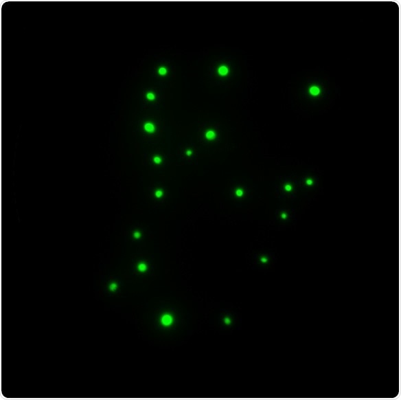
Figure 1. a) Green fluorescent colonies.
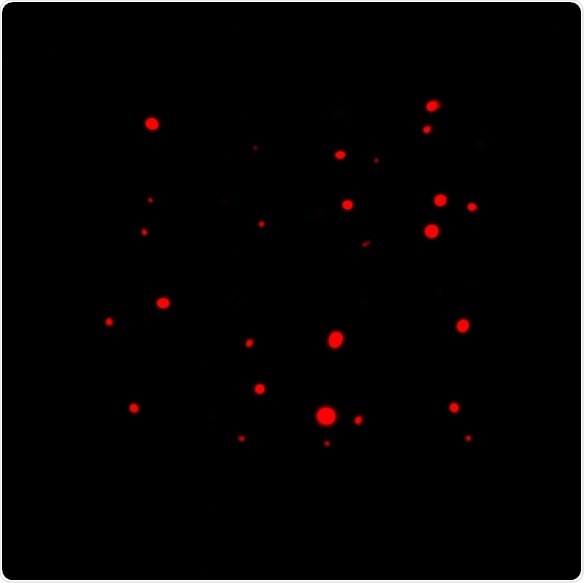
Figure 1. b) Red fluorescent colonies.
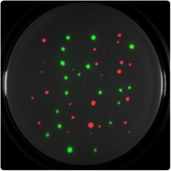
Figure 1. c) Composited green and red fluorescent colonies on a white light image.
These results confirm the ability of imaging systems such as the UVP GelSolo to increase visibility to all colonies, accurately and precisely detect various colony sizes, and supply highly sensitive image acquisition and analysis.
Automated colony count analysis was carried out on the green and red fluorescent colony images after imaging, using VisionWorks software. The green fluorescent plate contains a total of 19 colonies and the red fluorescent plate contains a total of 27 colonies according to the results shown in Figure 2A-B.
Compared to manual conventional techniques, VisionWorks imaging and analysis software supply a more accurate and intuitive colony counting approach. Colonies can be filtered by circularity and total pixel size after automatic colony counting for a more in-depth analysis. By using editing tools included in the software, researchers can also delete, add, split, or merge colonies.
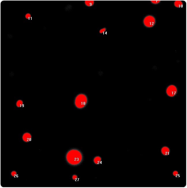
Figure 2. a) Green fluorescent colony count analysis.
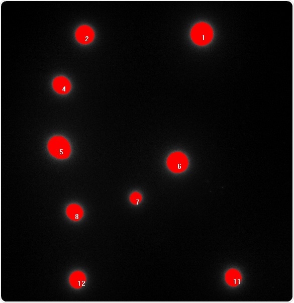
Figure 2. b) Red fluorescent colony count analysis.
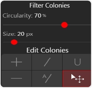
Figure 2. c) VisionWorks colony count editing and filter options.
Conclusion
As an easy-to-use system for colony documentation, the UVP GelSolo is designed for imaging and analysis without the requirement for any additional training. The straightforward features and user-friendly automation make the UVP GelSolo an ideal entry-level imager for multi-user laboratories, school laboratories, and practical training.
References
- Pious Thomas, Aparna C. Sekhar, Reshmi Upreti, Mohammad M. Mujawar, and Sadiq S. Pasha. Optimization of single plate-serial dilution spotting (SP-SDS) with sample anchoring as an assured method for bacterial and yeast cfu enumeration and single colony isolation from diverse samples. Biotechnol Rep (Amst). 2015 Dec; 8: 45–55.
- Mahboob Nemati, Aliasghar Hamidi, Solmaz Maleki Dizaj, Vahid Javaherzadeh, and Farzaneh Lotfipour. An overview on novel microbial determination methods in pharmaceutical and food quality control. Adv Pharm Bull. 2016 Sep; 6(3): 301–308.
About Analytik Jena US
Analytik Jena is a provider of instruments and products in the areas of analytical measuring technology and life science. Its portfolio includes the most modern analytical technology and complete systems for bioanalytical applications in the life science area.
Comprehensive laboratory software management and information systems (LIMS), service offerings, as well as device-specific consumables and disposables, such as reagents or plastic articles, complete the Group’s extensive range of products.
About life science
The Life Science product area demonstrates the biotechnological competence of Analytik Jena AG. We provide a wide product spectrum for automated total, as well as individual solutions for molecular diagnostics. Our products are focused to offer you a quality and the reproducibility of your laboratory results.
This will surely ease your daily work and speed up your work processes in a certain way. All together we support you through the complete process of the lab work. Besides we offer customized solutions and are able to adapt our products to your needs. Automated high-throughput screening systems for the pharmaceutical sector are also part of this segment’s extensive portfolio.
About analytical instrumentation
Analytik Jena has a long tradition in developing high-performance precision analytical systems which dates back to the inventions made by Ernst Abbe and Carl Zeiss. We have grown to become one of the most innovative manufacturers of analytical measuring technology worldwide.
Our business unit Analytical Instrumentation offers excellent competencies in the fields of optical spectroscopy, sum parameters and elemental analysis. Being proud of our core competency we grant all our customers a long-term warranty of 10 years for our high-performance optics.
About lab automation
With more than 25 years of market experience, Analytik Jena with its CyBio® Product Line is a leading provider for high quality liquid handling and automation technologies. In the pharmaceutical and life science industries, our products enjoy the highest reputation for precision, reliability, robustness and simplicity.
Moreover, the Automation Team designs, produces and installs fully automated systems tailored to our clients' application, throughput and capacity requirements. From stand-alone CyBio® Well up to fully customized robotic systems we handle your compounds, biomolecules and cells with great care.
Sponsored Content Policy: News-Medical.net publishes articles and related content that may be derived from sources where we have existing commercial relationships, provided such content adds value to the core editorial ethos of News-Medical.Net which is to educate and inform site visitors interested in medical research, science, medical devices and treatments.