Western blotting is an analytical method that is commonly used for the identification and quantification of specific proteins in a biological sample (Gallagher and Wiley, 2018).
Traditionally, a target protein is interrogated by antigen-specific primary antibodies that are then probed by secondary antibodies conjugated to either Alkaline Phosphatase (ALP) or Horseradish Peroxidase (HRP) and followed by chemiluminescent or colorimetric detection.
There continues to be growing interest in fluorescent protein (Western) blotting employing primary or secondary antibodies labeled with a fluorophore to perform non-enzymatic detection of protein expression because of the improved signal-to-noise and multiplexing capabilities.
Further benefits of fluorescent blotting include accurate quantitative analysis with broader dynamic range and high linearity in addition to excellent signal stability over time, reducing or eliminating the requirement to strip and reprobe.
Methods and materials
- Membrane: PVDF Millipore Immobilon-FL PVDF
- Imaging system: UVP ChemStudio
- Wash buffer: TTBS (Tween (0.1 % Tris Buffered Saline)
- Biotium’s TrueBlack WB blocking kit, Cat#23013 (blocking buffer and antibody diluents)
- Primary antibodies (0.1 μg/ml Primary Mouse or rabbit anti-tubulin)
- HeLa lysate. The total protein concentration is determined by BCA assay. The lysis buffer contains 20 mM Tris, 150 mM NaCl, 1 mM EDTA, 1 % TritonX-100, pH 7.5
- Secondary antibodies goat-∝-rabbit IgG – CF 680R or goat-∝-mouse IgG – CF 770®
Sample preparation
By utilizing a serial dilution of 10 μg, 7.5 μg, 5 μg, 2.5 μg, or 1 μg HeLa cell lysate protein was added into micro sample tubes to total 10 μl. According to manufacturer’s instructions (Biotium MnS Total Protein Prestain kit), lysate dilutions were pre-stained lysate for 30 minutes with CF680T or CF770T for 30 minutes.
Using a dry temp lock, loading buffer (4X LDS loading buffer)+ DTT was added to samples and heated for 3 minutes at 95 °C. Samples were loaded on a 4-12 % Bis-tris SDS-PAGE gel and separated at the constant voltage at 200V for 30 minutes.
Using an automated western blotting instrument according to manufacturer instructions, the separated proteins were then electrotransferred to a PVDF membrane at 20V for 1 hour.
Note. There are a variety of alternatives including semi-automated and manual strategies (Gallagher and Wiley, 2018).
Blots were processed according to the following sequence:
- Blocking, 4 °C 1 h
- Primary Ab binding, 4 °C 16 h
- Washing, 4 °C 5 x 10 min
- Secondary Ab binding, 4 °C 2 h
- Washing, 4 °C 5 x 10 min
Imaging
The UVP ChemStudio Imaging System and the integrated 660/787 nm lasers were utilized for fluorescent imaging. Images were composited with VisionWorks Touch software and were collected automatically. The processed blot was briefly positioned on the sample plate.
One-touch automated software set the optimal emission and excitation wavelengths, focus, and exposure for optimum signal without saturation and automatically pseudo-colored to show the two different wavelengths and compositing, in addition to saving the original raw data images. The original unaltered image was archived once collected and a copy was utilized for the image analysis.
Results and discussion
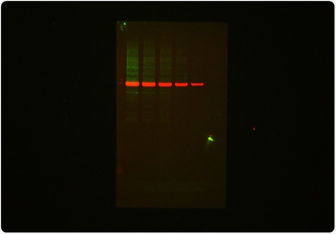
The multiplex imaging capabilities of the UVP ChemStudio Imaging System are shown in Figure 1, specifically separating out the signal of both Biotium CF680R and CF 770 fluorescence channels. Fluorescent western blotting applications provide a number of advantages over chromogenic or chemiluminescent visualization.
Most significantly, fluorescent labels enable multiplexing so that multiple proteins in a sample can be detected and analyzed simultaneously, on a single protein blot. In particular, for quantitative imaging, fluorescent labels provide extremely low background and a high signal-to-noise ratio.
The combination of 660 nm and 787 nm NIR excitation laser light sources with the UVP ChemStudio Imaging System with internal blue, green, red narrowband LEDs supplies a complete range of excitation light wavelengths.
It also provides fast, high-resolution image capture through the utilization of deeply cooled 815 8.1 mpx with NIR and QE enhanced 615 3.2 mpx cameras and f/0.95 low light lenses.

Figure 1. Protein blot stained with Biotium MnS Total Protein Prestain kit CF770T and Primary Rabbit anti-Tubulin/ goat-∝-rabbit IgG – CF 680R and visualized with the ChemStudio using the NIR 660/787 lasers set.
Imaging time
The UVP ChemStudio Imaging System supplies a significant advantage compared to laser scanning imaging systems where imaging times range from 3 to 5 minutes, as its imaging times are between 1 to 5 seconds for fluorescent Western blotting applications.
Conclusion
Fluorescent Western blot imaging with the UVP ChemStudio Imaging System is a quick and efficient process that allows researchers to achieve detection of multiple proteins on the same immunoblot to produce high resolution, publication-ready images.
References and Further Reading
Gallagher, S.R. and Wiley, E.A. Current Protocols: Essential Laboratory Techniques. Wiley, 2018
About Analytik Jena US
Analytik Jena is a provider of instruments and products in the areas of analytical measuring technology and life science. Its portfolio includes the most modern analytical technology and complete systems for bioanalytical applications in the life science area.
Comprehensive laboratory software management and information systems (LIMS), service offerings, as well as device-specific consumables and disposables, such as reagents or plastic articles, complete the Group’s extensive range of products.
About life science
The Life Science product area demonstrates the biotechnological competence of Analytik Jena AG. We provide a wide product spectrum for automated total, as well as individual solutions for molecular diagnostics. Our products are focused to offer you a quality and the reproducibility of your laboratory results.
This will surely ease your daily work and speed up your work processes in a certain way. All together we support you through the complete process of the lab work. Besides we offer customized solutions and are able to adapt our products to your needs. Automated high-throughput screening systems for the pharmaceutical sector are also part of this segment’s extensive portfolio.
About analytical instrumentation
Analytik Jena has a long tradition in developing high-performance precision analytical systems which dates back to the inventions made by Ernst Abbe and Carl Zeiss. We have grown to become one of the most innovative manufacturers of analytical measuring technology worldwide.
Our business unit Analytical Instrumentation offers excellent competencies in the fields of optical spectroscopy, sum parameters and elemental analysis. Being proud of our core competency we grant all our customers a long-term warranty of 10 years for our high-performance optics.
About lab automation
With more than 25 years of market experience, Analytik Jena with its CyBio® Product Line is a leading provider for high quality liquid handling and automation technologies. In the pharmaceutical and life science industries, our products enjoy the highest reputation for precision, reliability, robustness and simplicity.
Moreover, the Automation Team designs, produces and installs fully automated systems tailored to our clients' application, throughput and capacity requirements. From stand-alone CyBio® Well up to fully customized robotic systems we handle your compounds, biomolecules and cells with great care.
Sponsored Content Policy: News-Medical.net publishes articles and related content that may be derived from sources where we have existing commercial relationships, provided such content adds value to the core editorial ethos of News-Medical.Net which is to educate and inform site visitors interested in medical research, science, medical devices and treatments.