Gel electrophoresis acts as a confirmation that the correct nucleic acid product was produced in an experiment. Applications include checking successful PCR, nucleic acid fragment analysis after restriction enzyme treatment, or testing for genes that are associated with a disease of interest.
Agarose gel itself is a permeable matrix which enables charged nucleic acid fragments to migrate within the gel towards an opposite charge. The gel is placed in an electrophoresis chamber which is filled with a buffer that is able to conduct an electric current, this causes the nucleic acid to move.
Smaller fragments travel through the gel faster than larger fragments that are unable to move as far in the same amount of time. Ultimately creating a series of bands that can then be analyzed, this movement separates the fragments by size.
In order to visualize the bands in a gel, gel imaging is needed to detect fluorescent signals of nucleic acid binding dyes mixed into the gel or it is utilized to stain the gel post-electrophoresis.
As a widely employed method, in life sciences laboratories gel imaging systems and a number of binding dyes are commonplace. The UVP GelSolo is one such imaging system and it is built for high-resolution imaging optimized for maximum light sensitivity and quick capture speeds in long exposure or low-light applications.
With up to three UV wavelength options and an 8-48 mm manual lens, the UVP GelSolo supplies the flexibility to detect a wide scope of fluorescent wavelengths. Due to its low initial cost and adequate sensitivity1,2 ethidium bromide (EtBr) has been the predominant dye used for nucleic acid gel staining.
Yet, more and more laboratories have moved to safer alternatives, due to the additional costs and safety hazards associated with decontamination and waste disposal of EtBr. Developed by Biotium Inc, GelRed and GelGreen are two such alternatives to address safety concerns with EtBr while also enhancing dye sensitivity.
In this article, using an experimental simulation, a demonstration of how to gel imaging is attained in a research setting is shown. A silicone gel that contains plastic fluorescent rods will mimic a stained agarose gel.
The gel will be imaged via the UVP GelSolo system, demonstrating how the nucleic acid binding dyes GelRed and GelGreen look in a captured image. This article can be utilized as a teaching tool to supplement STEM curriculum by non-research institutions.
Using the same technique, further training can be done by those who may have access to imaging instruments to create an image a silicone gel.
Materials and methods
DNA binding dye simulation
GelRed is considered a nontoxic alternative dye which is more environmentally friendly than EtBr. However, the same excitation sources and emission filter can be used for both dyes (Figure 1.) as the spectrum of GelRed is similar to that of EtBr.
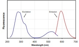
Figure 1. a) GelRed Excitation and Emission spectrum. (Source: GelRed: Biotium).
Figure 1. b) EtBr Excitation and Emission spectrum. (Source: EtBr: ThermoFisher).
GelRed dye bound DNA fragments are represented by red plastic fluorescent rods in the silicone gel in this experiment. GelGreen is another nontoxic dye option within a different wavelength range. Figure 2 shows the GelGreen spectrum.
GelGreen bound DNA fragments will be represented by plastic yellow-green fluorescent rods for this experiment. Both GelRed and GelGreen can be excited by 302 nm UV. The emission filter range employed for GelRed is 575-640 nm and for GelGreen is 513-557 nm.
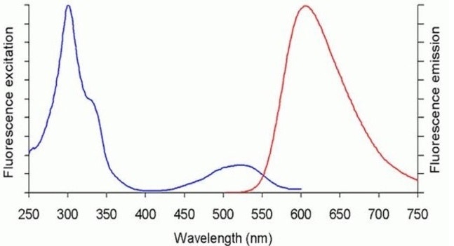
Figure 2. GelGreen spectrum. (Source: Biotium).
Silicone DNA gel
The silicone DNA Gel is created from semi-transparent silicone rubber. The red and yellow-green plastic fluorescent rods were lined up in columns on the surface of the solid silicone rubber. The second layer of liquid silicone was poured over the rods and then dried.
The resulting silicone gel is around 5 mm thick, this is the recommended thickness for standard agarose gels which are employed in DNA electrophoresis. There will be a lower resolution of the bands and higher background from staining if the agarose gel is thicker than 5 mm.
Silicone gel imaging
The silicone gels were imaged in the UVP GelSolo imaging system equipped with a 5.0-megapixel 315 camera and 8-48 mm f/1.2 manual zoom lens. 575-640 nm and 513-557 nm emission filters were placed in the filter slot located on top of the system to detect GelRed and GelGreen wavelengths. Table 1 shows the image capture data for each color dye.
| |
Exposure Time |
Filter |
| Red Fluorescent (GelRed) |
110ms |
575nm-640nm |
| Green Fluorescent (GelGreen) |
60ms |
513nm-557nm |
Image acquisition step-by-step procedure
- Turn on the UV transilluminator and switch the UV option to 302 nm.
- Double click to start VisionWorks software installed on the embedded touch computer.
- Choose the DNA Gel Electrophoresis application and begin a preview by clicking the Live View button.
- Adjust the zoom, aperture, and focus manually on the manual lens until the image looks clear and sharp.
- Click Capture to capture the image.
Results
Shown here by red and green fluorescent rods strategically placed in the silicone gels (Figure 3), the UVP GelSolo definitively detected both the GelRed and GelGreen wavelengths. The ability to image a wide range of wavelengths accommodates various binding dyes for gel imaging.
This feature enables the GelSolo to be versatile in its applications, whether for more in-depth genomic or proteomic studies or for the quick confirmation of product. Gel electrophoresis is a useful diagnostics tool in molecular biology, where nucleic acid fragments can be isolated, and an experiment continued successfully.
The sizes of the generated bands from the sample to be analyzed can be established by comparing sized bands from the simulated ladder on the left side of each gel in Figure 3. Fragments can also be excised from the gel and purified for further examination and manipulation, which is common in molecular cloning.

Figure 3. a) Silicone gel image with red pseudocolor representing GelRed bound DNA fragments.

Figure 3. b) Silicone gel with green pseudocolor representing GelGreen bound DNA fragments.
References and Further Reading
- Gallagher, SR and Wiley, EA. Current Protocols: Essential Laboratory Techniques. 2008.
- Armstrong, J and Schulz, J. 2008. Agarose Gel Electrophoresis. Curr. Protoc. Essential Lab. Tech. Unit 7.2
About Analytik Jena US
Analytik Jena is a provider of instruments and products in the areas of analytical measuring technology and life science. Its portfolio includes the most modern analytical technology and complete systems for bioanalytical applications in the life science area.
Comprehensive laboratory software management and information systems (LIMS), service offerings, as well as device-specific consumables and disposables, such as reagents or plastic articles, complete the Group’s extensive range of products.
About life science
The Life Science product area demonstrates the biotechnological competence of Analytik Jena AG. We provide a wide product spectrum for automated total, as well as individual solutions for molecular diagnostics. Our products are focused to offer you a quality and the reproducibility of your laboratory results.
This will surely ease your daily work and speed up your work processes in a certain way. All together we support you through the complete process of the lab work. Besides we offer customized solutions and are able to adapt our products to your needs. Automated high-throughput screening systems for the pharmaceutical sector are also part of this segment’s extensive portfolio.
About analytical instrumentation
Analytik Jena has a long tradition in developing high-performance precision analytical systems that date back to the inventions made by Ernst Abbe and Carl Zeiss. We have grown to become one of the most innovative manufacturers of analytical measuring technology worldwide.
Our business unit Analytical Instrumentation offers excellent competencies in the fields of optical spectroscopy, sum parameters, and elemental analysis. Being proud of our core competency we grant all our customers a long-term warranty of 10 years for our high-performance optics.
About lab automation
With more than 25 years of market experience, Analytik Jena with its CyBio® Product Line is a leading provider for high-quality liquid handling and automation technologies. In the pharmaceutical and life science industries, our products enjoy the highest reputation for precision, reliability, robustness and simplicity.
Moreover, the Automation Team designs, produces, and installs fully automated systems tailored to our clients' application, throughput, and capacity requirements. From stand-alone CyBio® Well up to fully customized robotic systems, we handle your compounds, biomolecules, and cells with great care.
Sponsored Content Policy: News-Medical.net publishes articles and related content that may be derived from sources where we have existing commercial relationships, provided such content adds value to the core editorial ethos of News-Medical.Net which is to educate and inform site visitors interested in medical research, science, medical devices and treatments.