Introduction
This protocol provides an overview for real time measurement of immune cell killing of adherent or non-adherent tumor cells. The highly flexible co-culture assay format is amenable to both antibody-dependent cell-mediated cytotoxicity (ADCC) and cytotoxic T-cell killing, two mechanisms of cell-mediated immune response.
This method integrates direct quantitation of tumor cell death with no-wash, mix-and-read protocols and makes use of either the IncuCyte® Caspase-3/7 reagent or the IncuCyte® Annexin V reagent.
The former is a DEVD caspase substrate which is cleaved during the apoptosis of target cells to release a green fluorescent DNA dye that stains the nuclear DNA, while the latter labels externalized phosphatidylserine (PS) moieties. Real-time automated detection and selective measurement of apoptotic tumor cell death is enabled by the IncuCyte® image analysis software.
Required materials
The following materials are used:
- IncuCyte® Annexin V Green Reagent (Sartorius Cat #4642) or
- IncuCyte® Caspase-3/7 Green Apoptosis Reagent (Sartorius Cat #4440) or
- IncuCyte® Caspase-3/7 GRedreen Apoptosis Reagent (Sartorius Cat #4704) or
- IncuCyte® Annexin V Red Reagent (Sartorius Cat #4641)
- Fibronectin (Sigma A7906), for non-adherent cells or
- Poly-L-ornithine (Sigma P4957), for non-adherent cells
- Flat bottom tissue culture plate (e.g., Corning 3595)
- Target cells of interest (non-adherent target cells have to be labeled with NucLight live-cell labeling reagent to allow tumor cell counting)
- IncuCyte® NucLight Green Lentivirus Reagent (Sartorius Cat # 4475)
- IncuCyte® NucLight Red Lentivirus Reagent (Sartorius Cat # 4476)
- 96-well microplate (e.g., Corning® 3595)
- Immune (effector) cells of interest
General guidelines
- To minimize the background of green fluorescence, a medium with low levels of riboflavin is recommended. RPMI and DMEM have high riboflavin (>0.2 mg/L). F12-K, EBM, and Eagles MEM have low riboflavin (<0.2 mg/L).
- After cell seeding, the plates should be placed at ambient temperature — 45 minutes for non-adherent cell lines and 15 minutes for adherent cell lines — to ensure uniform cell settling.
- Bubbles should be removed from all the wells by lightly squeezing a wash bottle (comprising of 70% to 100% of ethanol with the inner straw detached). This is done to blow vapor over the surface of all wells.
- Once the plate is placed in the IncuCyte® Live-Cell Analysis System, it should be allowed to warm to 37 °C for a period of 30 minutes before scanning.
Immune cell killing of adherent tumor cells protocol
Seed target cells
Seed tumor cells (100 μL/well, 1,000 to 3,000/well) into the 96-well plate.
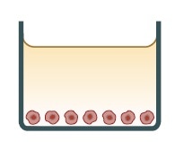
Optional: Target cells can be labeled with IncuCyte® NucLight Red live-cell labeling reagent (Sartorius 4476) to enable simultaneous tumor cell counting.
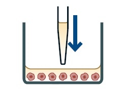
Treat cells
Aspirate the medium and add the Caspase-3/7 reagent or Annexin V reagent (50 μL/well) and desired treatments (50 μL/well) at 4x final assay concentrations.
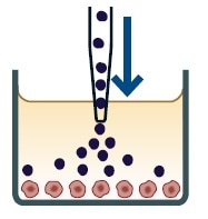
Add immune cells
Add your choice of immune cells (100 μL/well, 10,000 to 30,000/well) to a 96-well plate.
Day 0: Seed target cells
Seed target tumor cells (100 μL for each well) at a suitable density into a 96-well flat-bottom plate, so that the cell confluency becomes around 20% by day 1. For each tumor cell line used, the seeding density has to be optimized. But, it has been observed that 1,000 to 3,000 tumor cells for each well indicated practical starting points.
- The IncuCyte® Live-Cell Analysis System and confluence algorithm can be used for monitoring the target cell growth.
- Optional: To allow simultaneous real-time counting of viable tumor cells, NucLight Red live-cell labeling reagent (Catalog # 4475 or 4476) can be used to label target cells.
Day 1: Prepare apoptosis reagent and treatments
Apoptosis reagents must be diluted to ensure compatibility of apoptotic marker and target cell label, and treatments (such as cytokines, T cell stimuli, and antibodies) at 4 times final assay concentration in a preferred assay medium.
- If a Caspase-3/7 reagent is being used, it should be diluted to 20 μM (1:250 dilution) concentration, which is adequate for 50 μL per well.
- If Annexin V reagents are being using, Annexin V should be solubilized by adding 100 μL of PBS or complete medium. The reagents can be subsequently diluted in complete medium containing 1 mM CaCl2 for 1:50 dilution, which is adequate for 50 μL per well.
The cell plate should be removed from the incubator and the growth medium should be aspirated.
50 μL each of the prepared apoptosis reagent and treatments from the above first step should be added.
Note: add 50 μL of assay medium for treatment controls.
Add immune cells
The selected effector cells (such as PBMCs and T cells) should be counted, and this is followed by preparing a cell suspension at a density of 100,000 to 300,000 cells/mL (100 μL for each well, 10,000 to 30,000 cells/well). Testing of different target-to-effector cell ratios is recommended (e.g., 1:10, 1:5).
Note: Assay time can be reduced if the effector cells are pre-activated before adding them to the assay plate, but this may require a higher seeding density of target cells at the initial stage.
100 μL of effector cells should be seeded into the wells of the cell plate to obtain a final assay volume of 200 μL. The plates should be allowed to settle on a level surface at ambient temperature for a period of 30 minutes.
The assay plate must be placed into the IncuCyte® live-cell analysis system with repeat scanning scheduled for 24 hours:
- Channel selection: Phase Contrast + “Red” or “Green” based on the target cell label and the apoptosis reagent used
- Objective: 10x
- Scan type: Standard (2 images per well)
- Scan interval: Every 3 hours
Immune cell killing of non-adherent tumor cells protocol
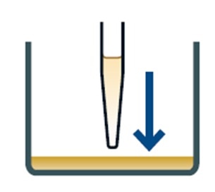
Coat plate
Coat plate surface to ensure even target cell coverage e.g. Poly-L-ornithine solution.
Prepare treatments
Prepare Annexin V reagent (50 μL/well) and desired treatments (50 μL/well) at 4x final assay concentrations.
Addition of target and effector cells
Add NucLight labeled target cells (50 μL/well, 10,000 to 20,000/well) and immune cells (50 μL/well, 100,000 to 200,000/well) to a 96-well plate.
Day 1: 1. Coat plate
Use a relevant coating matrix to coat a 96-well flat bottom plate. It is advisable to coat with 50 μL of 5 μg/mL fibronectin or 0.01% poly-L-ornithine solution diluted in 0.1% BSA.
Coat the plates at ambient temperature for 1 hour, remove the solution from the wells, and then allow the plates to dry for 30 to 60 minutes before adding the cells.
Before the assay, the type of coating to be used should be determined for target cells of interest.
2. Reagent and treatment preparation
The following reagents should be prepared in medium:
- Test materials (such as cytokines, antibodies, and T cell stimuli; 50 μL per well, prepared at 4 times final assay concentration)
- Apoptosis detection reagent (make sure to check the compatibility of apoptotic marker and cell label), IncuCyte® Annexin V Reagent (Cat # 4641 or 4642): Annexin V is solubilized by adding 100 μL of PBS or complete medium and the reagents can be diluted in complete medium containing 1 mM CaCl2 for 1:50 dilution (4 times final assay concentration, 50 μL for each well)
All the prepared reagents should be added to the assay plate to acquire 100 μL per well.
Note: While immune cell killing of target cells can be detected using either the Caspase-3/7 or IncuCyte® Annexin reagent, the Annexin V reagent is recommended for non-adherent target cells.
Effector cells and non-adherent target cells can exhibit similar nuclear sizes, removing the need for size filters to eliminate Caspase-3/7 labeled effector nuclei from the analysis. Moreover, increased levels of Caspase-3/7 activity were observed in certain non-adherent cell types, especially at higher confluency.
This can impede with the interpretation of target cell death driven by immune cells. Typically, fewer effector cells are labeled by the Annexin V reagent resulting in lower non-specific background.
All the prepared reagents should be added to the assay plate to obtain 100 μL per well.
3. Add immune cells
Labeled target cells have to be counted and a cell suspension should be prepared at a density of 40,000 – 80,000 cells/mL (seed 50 μL per well, 10,000 to 20,000 cells/well). NucLight Green or Red live-cell labeling reagent (Cat # 4476 or 4475) can be used to label the target cells to achieve real-time counting of viable tumor cells simultaneously.
The selected effector cells (such as PBMCs, T cells) should be counted and a cell suspension at a density of 400,000 to 800,000 cells/mL (50 μL per well, 100,000 to 200,000 cells/well) should be prepared. It is recommended that different target-to-effector cell ratios are tested (e.g. 1:5, 1:10).
Note: The duration of the assay can be reduced by pre-activating the effector cells prior to adding them to the assay plate, but a higher initial seeding density of target cells may be required for this.
Target and effector cells should be added to the assay plate to obtain a final assay volume of 200 μL. The plates should be allowed to settle on level surface at ambient temperature for a period of of 30 minutes.
The assay plate should be placed into the IncuCyte® instrument with repeat scanning scheduled for 24 hours.
- Channel selection: Phase Contrast + “Red” and “Green”
- Objective: 4x
- Scan type: Standard (2 images per well)
- Scan interval: Every 2 to 3 hours
Related products and applications
A complete range of cell health and fluorescent nuclear labeling reagents are supplied by Sartorius. These can be used with the IncuCyte® Live-Cell Analysis System to allow multiplexed measurements of proliferation, apoptosis and cytotoxicity. A complete range of cell health applications is available to suit specific experimental requirements.
|
Product
|
Cat No.
|
Amount
|
|
IncuCyte® NucLight Green Lentivirus Reagent (EF-1 α, Puro) for nuclear labeling
|
4624
|
0.2 mL
|
|
IncuCyte® NucLight Red Lentivirus Reagent (EF-1 α, Puro) for nuclear labeling
|
4625
|
0.2 mL
|
|
IncuCyte® NucLight Green Lentivirus Reagent (EF-1 α, Bleo) for nuclear labeling
|
4626
|
0.2 mL
|
|
IncuCyte® NucLight Red Lentivirus Reagent (EF-1 α, Bleo) for nuclear labeling
|
4627
|
0.2 mL
|
|
IncuCyte® NucLight Green Lentivirus Reagent (EF-1 α, Puro) for nuclear labeling
|
4475
|
0.6 mL
|
|
IncuCyte® NucLight Red Lentivirus Reagent (EF-1 α, Puro) for nuclear labeling
|
4476
|
0.6 mL
|
|
IncuCyte® NucLight Green Lentivirus Reagent (EF-1 α, Bleo) for nuclear labeling
|
4477
|
0.6 mL
|
|
IncuCyte® NucLight Red Lentivirus Reagent (EF-1 α, Bleo) for nuclear labeling
|
4478
|
0.6 mL
|
|
IncuCyte® Cytotox Red Reagent for counting dead cells
|
4632
|
5 μL x 5
|
|
IncuCyte® Cytotox Green Reagent for counting dead cells
|
4633
|
5 μL x 5
|
|
IncuCyte® Annexin V Red Reagent for apoptosis
|
4641
|
100 tests
|
|
IncuCyte® Annexin V Green Reagent for apoptosis
|
4642
|
100 tests
|
|
IncuCyte® Caspase-3/7 Green Reagent for apoptosis
|
4440
|
20 μL
|
|
IncuCyte® Caspase-3/7 Red Reagent for apoptosis
|
4704
|
20 μL
|
 Sartorius
Sartorius is a leading international pharmaceutical and laboratory equipment supplier. With our innovative products and services, we are helping our customers across the entire globe to implement their complex and quality-critical biomanufacturing and laboratory processes reliably and economically.
The Group companies are united under the roof of Sartorius AG, which is listed on the Frankfurt Stock Exchange and holds the majority stake in Sartorius Stedim Biotech S.A. Quoted on the Paris Stock Exchange, this subgroup is comprised mainly of the Bioprocess Solutions Division.
Innovative Technologies Enable Medical Progress
A growing number of medications are biopharmaceuticals. These are produced using living cells in complex, lengthy and expensive procedures. The Bioprocess Solutions Division provides the essential products and technologies to accomplish this.
In fact, Sartorius has been pioneering and setting the standards for single-use products that are currently used throughout all biopharmaceutical manufacturing processes.
Making Lab Life Easier
Lab work is complex and demanding: Despite repetitive analytical routines, lab staff must perform each step in a highly concentrated and careful way for accurate results.
The Lab Products and Services Division helps lab personnel excel because its products, such as laboratory balances, pipettes and lab consumables, minimize human error, simplify workflows and reduce physical workloads
Sponsored Content Policy: News-Medical.net publishes articles and related content that may be derived from sources where we have existing commercial relationships, provided such content adds value to the core editorial ethos of News-Medical.Net which is to educate and inform site visitors interested in medical research, science, medical devices and treatments.