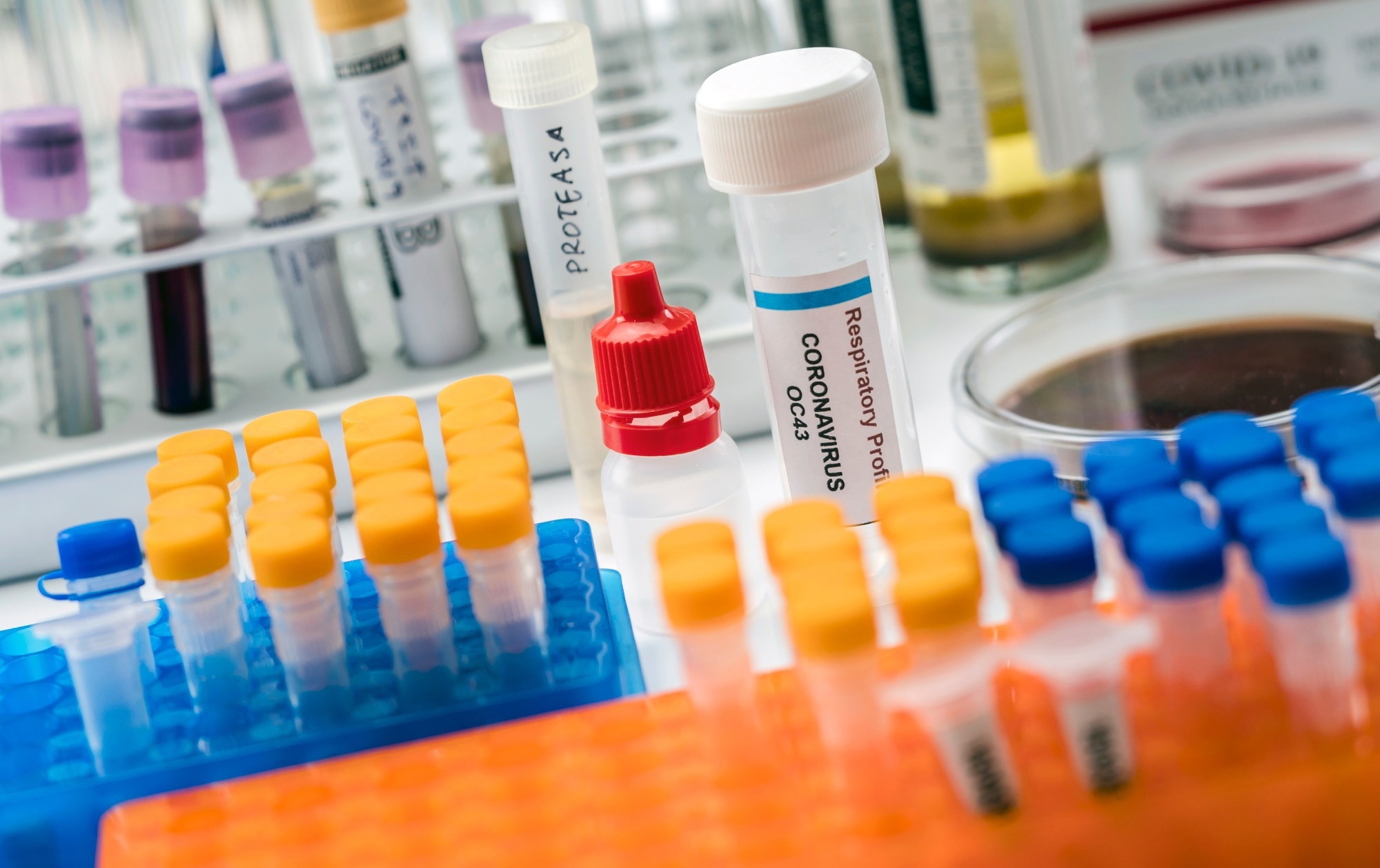
In addition, they examined the effects of trypsin on five clinical samples comprising SARS-CoV-2 Delta variants in the presence of the culture supernatant of anaerobic bacteria, e.g., Fusobacterium necrophorum.
 Study: Activation of SARS-CoV-2 by trypsin-like proteases in the clinical specimens of patients with COVID-19. Image Credit: felipecaparros/Shutterstock.com
Background
Most respiratory viruses, including SARS-CoV, influenza virus, and SARS-CoV-2, need proteases to infect and proliferate in epithelial cells of upper respiratory tract (URT) airways.
Tryptase Clara cleaves the hemagglutinin of the influenza virus near airway epithelial cells, a step crucial for influenza infection. Likewise, SARS-CoV-2 infects by endocytosis using the angiotensin-converting enzyme 2 (ACE2), a protease.
Furthermore, another cellular protease, transmembrane protease, serine 2 (TMPRSS2) enhances the infectivity, i.e., the yield of SARS-CoV-2 virions by cell membrane fusion inside the host.
Studies have also reported that staphylococcal proteases and proteases from oral bacteria increase viral infectivity in the URT. Thus, having a clean oral cavity clean reduces the number of proteases derived from indigenous bacteria, which, in turn, helps prevent infection from several types of viruses.
About the study
In the present study, researchers used clinical samples positive for SARS-CoV-2 from COVID-19 patients at the Toyama Institute of Health in Japan. They used next-generation genome sequencing to identify Delta and Omicron variant specimens.
Further, the team generated pseudotyped vesicular stomatitis viruses (VSVs) bearing spike (S) protein of SARS-CoV-2 (SARS-CoV-2pv) for the trypsinization experiments. Specifically, they examined the effect of trypsin on SARS-CoV-2pv or VSVpv by exposing them to various concentrations of trypsin in Dulbecco’s modified Eagle’s medium (DMEM) for five minutes at 37 °C.
The team used reverse transcriptase-polymerase chain reaction (RT-qPCR) to quantify virus titer in the cell culture supernatants of VeroE6 cells. Furthermore, the researchers examined syncytium formation after trypsin treatment in Vero E6 cells and whether oral anaerobic bacteria increased SARS-CoV-2 infectivity.
To this end, they used Prevotella melaninogenica, P. intermedia, and F. necrophorum, three species of anaerobic bacteria from the Japan Collection of Microorganisms.
Results
As its effects on other animal and human coronaviruses, trypsin increased SARS-CoV-2 entry and SARS-CoV-2pv infectivity. Trypsin increased the infectivity of the Wuhan, Alpha, and Delta variants but not the SARS-CoV-2pv Omicron variant in a concentration-dependent manner. For the former three SARS-CoV-2pv variants, trypsin treatment at 800 μg/mL concentration increased the infectivity by up to ~ 10-fold.
Treatment with trypsin also increased the infectivity of SARS-CoV-2 present in clinical samples derived from the nasal/oral cavities of COVID-19 patients. The authors noted an increase of up to 36,000-fold in infectivity of the Delta variant after trypsin treatment, whereas it increased to a meager few dozen-fold for Omicron.
Another intriguing observation of this study was that the trypsin-induced effect on SARS-CoV-2 infectivity became apparent only after the virus bound its cellular receptor ACE2 on the host cells. Thus, trypsin pre-treatment did not increase the infectivity of SARS-CoV-2pv variants.
In SARS-CoV-2, syncytium formation also plays a role in the pathogenesis of severe COVID-19. Cellular membrane fusion activity is lower in Omicron than in Delta as it uses a TMPRSS2-independent entry pathway.
Thus, even though trypsin-like proteases activate cell membrane fusion, the enhancement of infection by trypsin treatment in the Omicron variant was lower than in Delta.
Studies have shown that the nasopharyngeal microbiome of COVID-19 patients is distinct from healthy people, even though they also have several types of commensal bacteria in their oral cavities. COVID-19 patients with severe illness had a marked abundance of operational taxonomic units (OTUs) classified as Prevotella.
In this study, culture supernatants of F. necrophorum but not P. melaninogenica and P. intermedia increased SARS-CoV-2 infectivity in the nasal cavity, thus, raising the need to investigate the presence, magnitude, and type of the protease in the culture supernatants.
Moreover, studies should investigate and elucidate the interaction mechanisms between the oral microbiome and SARS-CoV-2.
Conclusions
To summarize, reducing components, especially proteases derived from oral indigenous bacteria, possible by daily cleaning the oral cavity is the key to preventing SARS-CoV-2 infection.
More importantly, people at an increased risk of severe COVID-19 must maintain oral hygiene, including chemical plaque control.