Small extracellular vesicles (sEVs) are excellent nanoscale protein carriers. For example, in a recent study, researchers used sEVs as a decoy for severe acute respiratory syndrome coronavirus 2 (SARS-CoV-2) by displaying the virus's host receptor on the surface of the sEV.
Characterizing these engineered sEVs, the researchers demonstrated that the soluble angiotensin-converting enzyme 2 (sACE-2) loaded sEV successfully binds and thus inhibits the entry of wild-type SARS-CoV-2 as well as its variants, protecting against SARS-CoV-2 infection. They also demonstrated the therapeutic efficacy of these engineered sEVs in vivo.
This study is a recent short communication in the Journal of Extracellular Vesicles.
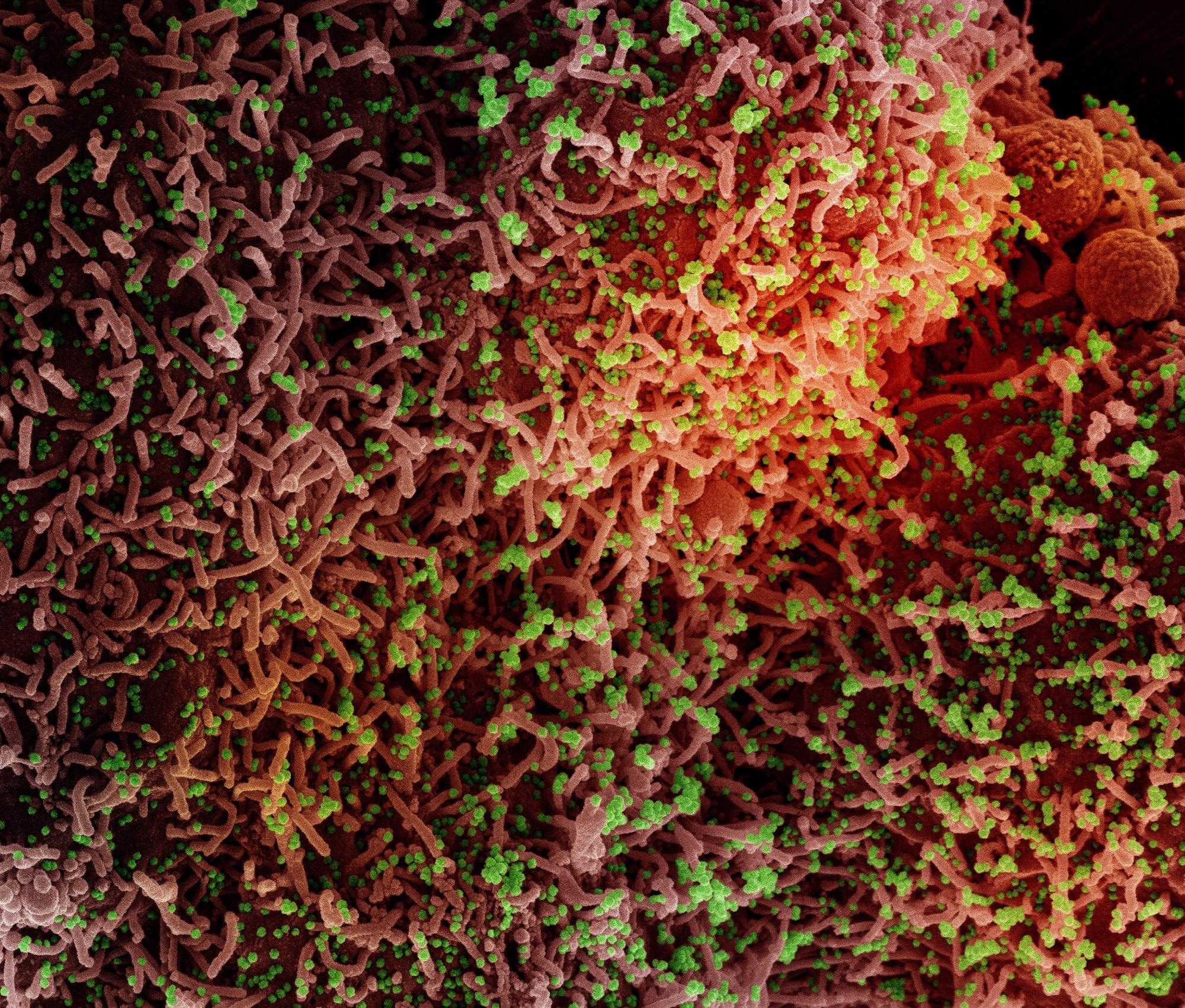
Study: Engineered small extracellular vesicles displaying ACE2 variants on the surface protect against SARS-CoV-2 infection. Image Credit: NIAID
Introduction
With the emerging of new variants and increasing cases of infections and hospitalizations, finding an effective therapeutic solution is necessary to control the ongoing COVID-19 pandemic caused by the SARS-CoV-2.
To this end, scientists are exploring the possibility of neutralizing the virus by utilizing its binding affinity. The viral spike (S) protein binds to the host ACE-2 receptor to gain entry into the human host cells. ACE2 is present on cells' surface in the lungs, intestine, kidneys.
Recent studies have reported that the human recombinant soluble ACE-2 (sACE-2) is capable of inhibiting SARS-CoV-2 infection in kidney organoids.
Incentivizing in this direction, in the present study, the researchers employed sEVs as cargo carriers of sACE-2 and a protein CD9 as a scaffold to decoy SARS-CoV-2 - into binding with the engineered sEVs instead of entering cells and causing infection.
sEVs are protein carriers, ranging 50–200 nm in diameter. It includes exosomes and microvesicles. They can transfer diverse cargos, including proteins, lipids, and RNAs, facilitating cell-to-cell communication and other diverse functions. sEVs are exploited as therapeutic transporters.
CD9 is a protein - one of the essential exosomal surface proteins, belonging to the tetraspanin superfamily. The tetraspanin proteins localize subcellular followed by cytosolic puncta and membrane localization. The CD9 has four transmembrane regions and two extracellular loops. It is found that CD9 overexpression enhances the production rate of sEVs.
In this study, the researchers have used CD9 as a scaffold to display active sACE-2 on the surface of the sEVs. The sEVs carried the sACE-2 of the wild type and its variants using it as an extracellular decoy and neutralized the S protein of SARS-CoV-2.
Study design and findings
For utilizing the CD9 as a scaffold, the researchers deleted the transmembrane region 4 to generate a truncated form of the CD9 (CD9ΔTM4). The C terminus end of this CD9ΔTM4 was conjugated with the sACE2 to have a functional sACE2 on the surface of the sEVs (CD9ΔTM4-sACE2).
Previous reports observed that the mutations in ACE2 enhanced the binding to the viral S protein. Similarly, with deep mutagenesis, the researchers in the present study used mutated forms of sACE2 for higher binding affinity for the S protein. They utilized the CD9ΔTM4-sACE2(WT) scaffolds and also used two of the variants, sACE2.v1 and sACE2.v2, and characterized these according to the MISEV2018 (Minimal Information for Studies of Extracellular Vesicles 2018) guidelines. The analysis determined their particles size and concentration and the high purity of EVs despite overexpression of sACE2.
Performing an ACE2:S protein inhibitor screening assay and immunoprecipitation (IP) assays to find the binding affinities, the researchers found that the engineered sEVs had super affinity for the S protein.
These sEVs had the capacity to neutralize SARS-CoV-2 pseudovirus bearing the viral envelope S protein, containing the Beta and the Delta variants with mutations. Further, the sEVs-mediated protein delivery conferred more superior activity in neutralization than the recombinant protein. Thus, the results demonstrated that these sEVs exhibited cross-reactive neutralization against the wild-type and mutant S protein pseudotyped virus.
Comparing sEVs displaying sACE-2 from variants vs wild-type, the researchers found that the sACE2 variants harboring sEVs showed superior antiviral efficacy.
More importantly, the engineered sEVs must be capable of suppressing the SARS-CoV-2 infectivity to come to potential clinical aid. The researchers tested against the authentic wild-type SARS-CoV-2 and its Delta variant infection in Vero E6 cells and reported high efficacy in inhibition of the infection.
Likewise, the researchers also tested the engineered sEVs to protect against SARS-CoV-2 infection in K-hACE mice. These are transgenic mice expressing human ACE2 under cytokeratin 18 gene promoter. Because the sACE2.v1 sEVs exhibited the best efficacy in vitro experiments, the researchers tested in mice. Observing the mice for weight loss, viral burden, and cytokine profile, the researchers concluded that sACE2.v1 conferred protection against SARS-CoV-2 infection in vivo.
Conclusions
This study demonstrated that the engineered sEVs - on CD9ΔTM4 scaffolds - displaying sACE2, successfully inhibited interactions of S protein with ACE2 and neutralized pseudovirus, authentic wild-type, and Delta variant SARS-CoV-2 entry, via in vitro and in vivo experiments. The results here strongly support the potential of engineered sEVs as an efficient platform for the delivery of therapeutics.
Journal reference:
- Kim, H. K., Cho, J., Kim, E., Kim, J., Yang, J.-S., Kim, K.-C., Lee, J.-Y., Shin, Y., Palomera, L. F., Park, J., Baek, S. H., Bae, H.-G., Cho, Y., Han, J., Sul, J. H., Lee, J. Park, J. H., Cho, Y. W., Lee, W., & Jo, D.-G. (2022). Engineered small extracellular vesicles displaying ACE2 variants on the surface protect against SARS-CoV-2 infection. Journal of Extracellular Vesicles, 11, e12179. https://doi.org/10.1002/jev2.12179, https://onlinelibrary.wiley.com/doi/10.1002/jev2.12179
 Large Swedish study finds COVID-19 vaccination unrelated to fertility or childbirth rates