Its identification will help enhance the understanding of protective immunity in the human IFN response.
The study used genome-wide Clustered Regularly Interspaced Short Palindromic Repeats (CRISPR)–CRISPR-associated protein 9 screens of human lung epithelial cells and hepatocytes ectopically expressing angiotensin-converting enzyme 2 (ACE2) to study the function of PLSCR1 against live SARS-CoV-2 before and after stimulation with IFNγ.
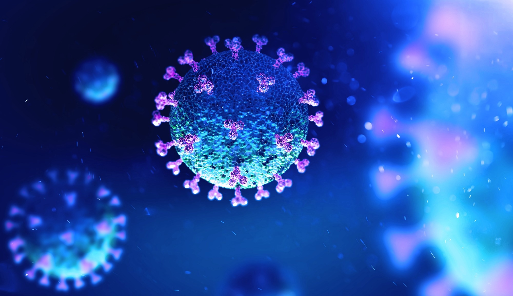
 Study: PLSCR1 is a cell-autonomous defence factor against SARS-CoV-2 infection. Image Credit: AndriiVodolazhskyi/Shutterstock.com
Background
In many plants, bacteria, and animals, including humans, cell-autonomous immunity helps combat viral and other infections, e.g., Shigella flexneri and Mycobacterium tuberculosis. IFNγ, a type II cytokine, is a well-recognized agent of mobilization of cell-autonomous immunity in most nucleated cells.
Thus, studies have found that increased expression of type I and III IFNs (IFNα\β and IFNλ) confers protection against coronavirus disease 2019 (COVID-19) in adults and children.
On the contrary, genetic lesions in IFN signaling and type I and II IFN autoantibodies are responsible for up to 20% of critical COVID-19 cases. T cells also recognize COVID-19 vaccines and SARS-CoV-2 variants of concern (VOCs) by secreting IFNγ.
So far, the research focus has been utilizing neutralizing antibodies (nAbs) as COVID-19 therapeutics. Recent evidence that IFNγ therapy rescued immunocompromised patients with COVID-19 for whom convalescent plasma or remdesivir treatment failed prompted the research community to investigate whether IFNγ could orchestrate anti-SARS-CoV-2 defense.
About the study
In the present study, researchers used human Huh7.5 hepatoma and A549-ACE2 cells (mimicking host-cell targets within the respiratory tract) to test IFNγ potency in restricting live SARS-CoV-2 infection, which they confirmed using CRISPR–Cas9 engineering.
Next, they used genome-wide loss-of-function (LoF) screens to identify which IFNγ-induced restriction factors conferred this effect against SARS-CoV-2.
The researchers used a version 2.0 GeCKO single-guide ribonucleic acid (sgRNA) library to transduce each cell type, and then, they performed puromycin selection to ensure stable integration. This library had 122,441 sgRNAs spanning 19,050 genes.
They used SARS-CoV-2 expressing mNeonGreen (mNG) to infect sgRNA-integrated cells treated with recombinant human IFNγ. Then, with the help of fluorescence-activated cell sorting (FACS), the researchers separated SARS-CoV-2-infected cells into mNGhigh and mNGlow populations, where the former were permissive while the latter was restrictive. Next, they performed next-generation sequencing of sgRNA frequencies.
In addition, the team identified PLSCR1 identified which step of the SARS-CoV-2 life cycle and how it blocked coronavirus invasion via a cathepsin-dependent endosomal fusion pathway or transmembrane serine protease 2 (TMPRSS2)-dependent cell-surface fusion pathway.
Further, the team used a split-NanoLuc-reporter-based assay to test virus–endosome fusion in the host cell cytosol. Furthermore, they combined nanoscale imaging with protein mutagenesis to identify the membrane determinants required to distinguish how PLSCR1 impeded SARS-CoV-2 fusion.
Results and conclusion
The current study had several important findings. First, the authors found that after exposure to recombinant human type II IFNγ, Huh7.5 cells restricted SARS-CoV-2 in a signal transducer and activator of transcription-1 (STAT1)-dependent manner.
Second, IFNγ-induced PLSCR1 restricted ancestral SARS-CoV-2 strain, USA-WA1/2020; however, it was also effective against the Delta and Omicron BA.1 VOCs. Its antiviral activity remained robust even against other highly pathogenic coronaviruses, having bats and mice as hosts.
Third, in PLSCR1-knockout cells, SARS-CoV-2 spike(S)-mediated cell–cell fusion increased significantly, while PLSCR1 overexpression reduced this response. These observations confirmed that PLSCR1 directly prevented membrane fusion triggered by the SARS-CoV-2 S protein.
Likewise, in A549-ACE2 cells, PLSCR1-enriched foci appeared within 30 minutes of viral infection and completely overlapped viral particles detected by anti-S antibodies. PLSCR1 acted against SARS-CoV-2 entry and targeted the S-mediated viral entry.
Intriguingly, it interfered with the uptake of SARS-CoV-2 in both the endocytic and the TMPRSS2-dependent fusion routes before viral RNA release into the host-cell cytosol, i.e., at a late entry step. Thus, PLSCR1 mutations could make people highly prone to severe COVID-19, as confirmed by a recent report.
Structural modeling predicted that PLSCR1 had a flexible N-terminal domain and 12-stranded membrane β-barrel that buried the C-terminal hydrophobic helix.
Once docked to the target plasma membrane by palmitoylation, PLSCR1 used its C-terminal β-barrel to disrupt the fusion of the virus with the host cell, referred to as fusogenic blockade, as revealed by nanosecond molecular dynamics simulation (MDS) analysis.
PLSCR1 likely occupied plasma-membrane microdomains used by coronaviruses to direct distinct subpopulations of SARS-CoV-2 vesicles. Given its palmitoyl anchorage, it operated on high- and low-curvature (endosome and plasma membrane) membranes to inhibit S-mediated fusion.
Conclusion
Overall, this study provided a mechanistic framework for PLSCR1 in blocking SARS-CoV-2-S-mediated fusion by virulent coronaviruses.
It highlights the PLSCR1 β-barrel as a major structural determinant of cell-autonomous immunity to the large coronavirus family.