X-ray photoelectron spectroscopy (XPS) is widely used for characterizing the uppermost 10 nm of a materials surface. Also called electron spectroscopy for chemical analysis (ESCA), the analytical technique measures photoelectrons ejected from a material under irradiation by monochromated Al Kα X-rays. XPS provides quantitative elemental and chemical state information from the materials surface. The chemistry of a device's surface can be critical in its with it's interaction with its environment and therefore its application. The AXIS Supra+ can be used to characterise surfaces as diverse as drug-loaded resorbable coatings to anti-fouling surface coatings.
The AXIS Supra+—Optimized Performance
Kratos Analytical’s next-generation AXIS Supra+ ensures ease of use and high sample throughput by integrating industry-leading imaging and spectroscopic capabilities with automation. Unparalleled large-area spectroscopic performance enables users to acquire photoelectron spectra. Fast XPS imaging with high spatial resolution exposes the lateral distribution of surface chemistry and helps in additional characterization by selected area analysis.
The AXIS Supra+ provides completely automated sample handling through computer control. At the time of analysis, a sample holder can be transferred and exchanged without operator assistance.
Capabilities of the AXIS Supra+ : Large-Area, High-Sensitivity XPS
Kratos Analytical’s AXIS Supra+ has been designed for chemical-state XPS. High transmission electron optics along with an efficient collection of photoelectrons ensures unrivaled resolution and sensitivity at large analysis areas.
Main spectroscopic attributes:
- Rapid data acquisition
- Easy identification of light elements
- Exceptional signal-to-noise ratio, even at trace concentrations
High-Energy Resolution
The major requirement of any spectrometer is the best possible energy resolution. As such, high-energy resolution, where the spectrometer does not play a part in the widening of the photoemission peaks, is important to accurately measure the slight chemical shifts. Kratos Analytical’s AXIS Supra+ uses a monochromated Al Kα X-ray source as well as enhanced electron optics, guaranteeing exceptional chemical resolution.
Benefits of the high spectral energy resolution are given below:
- Guaranteed energy resolution on both conducting and insulating samples
- Accurate detection of chemical shifts
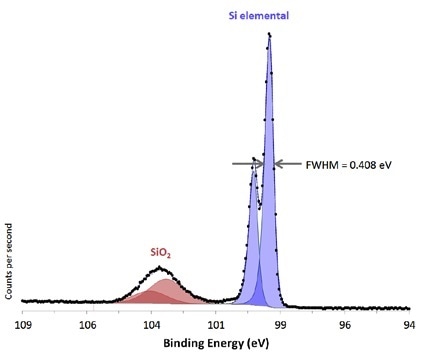
Si 2p region from native oxide on Si substrate acquired from large area with high-energy resolution.
Small Spot, Selected Area Spectroscopy
Enhanced performance of the AXIS Supra+ ensures high sensitivity in small spot modes as small as 15 μm diameter. This is realized by matching X-ray illumination with the chosen analysis area. An optional brite-X monochromatic source can also be specified for applications requiring extremely high sensitivity performance from minute selected regions.
Main attributes of the selected area spectroscopy are listed below:
- Either achromatic or monochromatic X-ray source can be utilized to acquire the chosen area spectra
- Improved X-ray illumination for better performance of the selected area
- Predefined small-spot analysis, 110 μm, 55 μm, 27 μm and 15 μm
- Optional μ-boost acquisition mode for very high sensitivity at small area mode
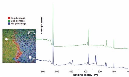
XPS image used to define a position for 27-μm selected area survey spectra.
Fast Parallel Imaging
XPS imaging is used to determine the lateral distribution of either chemistry or elements at the surface. Rapid and high-spatial-resolution parallel images are acquired by the AXIS Supra+ instrument.
Furthermore, parallel imaging can be integrated with stage movements to get a “stitched” image, capable of producing images over several millimeters with a spatial resolution of several microns.
Parallel imaging provides the following capabilities:
- Spectromicroscopy—Spectra can be easily acquired from image datasets, enabling a spectrum at every pixel
- High-energy resolution, chemical-state imaging
- Maximum spatial resolution of 1 μm at highest level of magnification
- Quantitative imaging—the special spherical mirror analyzer together with the delay-line detector can offer quantitative chemical-state images
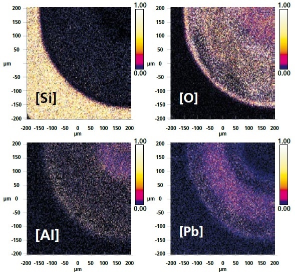
Quantitative images where the color scale represents a relative amount of each element present at the surface.
Versatile ESCApe Software for Acquisition and Processing
The versatile ESCApe software has been developed to make sure that users’ interaction with the spectrometer is made as easy as possible, combining acquisition and processing to completely manipulate the hardware automation. An example of this is the group array analysis capability within the ESCApe software. Through this functionality, users can drag an area over a sample and define a range of analysis points from which spectra are obtained.
Automated identification and quantification of peaks make it easy to produce a color concentration map of detected elements over the surface of the sample.
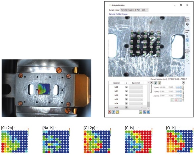
Group array analysis of an inhomogeneous distribution of Cu-nanoparticles on a graphite substrate. The color scale used to represent relative elemental concentration.
Multi-Technique Capability
Optional excitation sources comprise a high energy Ag Lα monochromated X-ray source, an enhanced helium discharge lamp for ultraviolet photoemission spectroscopy (UPS), as well as an achromatic Al/Mg X-ray source.
The inclusion of a field-emission electron source also adds scanning auger mapping (SAM), auger electron spectroscopy (AES), and secondary electron microscopy (SEM) capability to the instrument. Additional methods like these are coincident with the XPS analysis position, providing a complementary understanding of the sample. Significantly, the inclusion of these methods does not influence the performance of the industry-leading XPS.
Options like surface modification and sample preparation can be fitted on the introduction chamber, also called Flexi-lock. Such options comprise sample heat and cool, broad spot ion source, air-sensitive sample transporter, crystal cleaver, and glove box.
A dedicated UHV setting for surface science research can be provided by configuring a third chamber. Usual configuration outfits this third chamber with a manual stage as well as optional characterization methods like quadrupole secondary ion mass spectrometry (SIMS), inverse photoemission spectroscopy (IPES), and low-energy electron diffraction (LEED).
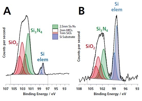
Si 2p spectra acquired from a thin-film multilayer sample (courtesy of IMEC) shown in the schematic figure using (A) monochromated Al Kα radiation and (B) monochromated Ag Lα. The greater information depth of the Ag Lα excited Si 2p spectrum is demonstrated by the larger Si elemental substrate component.
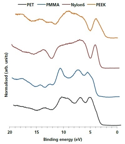
He II excited UPS valence band spectra of four common polymers.
Into the Bulk—Ion Sputter Sources
The AXIS Supra+ may be configured with either the Minibeam 6 Arn+ gas cluster ion source (GCIS) or the Minibeam 4, high-flux Ar+ monatomic ion source . These two ion sources are controlled through the ESCApe acquisition software for depth profile experiments or sputter cleaning. Gas handling is completely automated for sputter profiling and contains pump/purge sequences to enable the transition to helium gas for ion scattering spectroscopy (ISS).
The Minibeam 6 is a multi-mode ion source that can work in Ar+/He+ monatomic or Arn+ cluster modes, as required. Gas cluster ion sources developed recently pave the way for depth profiling organic materials with retention of chemistry across the profile. Concentration depth profiles can be aquired from samples as diverse as tablets or drug release coatings on polymeric stents.
The Minibeam 4 ion source is provided where cluster ion depth profiling capability is not required. This ion source works with continuously variable beam energies ranging from 500 eV to 4 keV. The design generates a high ion flux at low energy for better interface resolution and, at the same time, retains a high sputter rate.
- An option of monatomic or multi-mode gas cluster ion sources
- Both sources can operate with He+ ions for optional ISS mode
- Automated introduction of gas and regulation of pressure
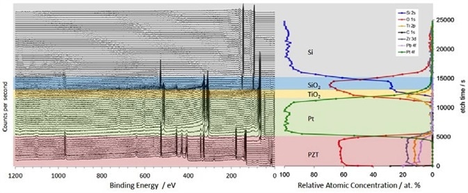
Sputter depth profile survey spectra (left) and elemental concentration as a function of etch time (right) through a PZT/Pt/TiO2/SiO2/Si multilayer sample using 20 keV Ar500+ ions.
Unrivaled Instrument Automation
Sample handling is completely automated in the AXIS Supra+, making it different from other spectrometers. The automatic sample exchange at the conclusion of an experiment enables continuous throughput of samples.
Automation extends to the monochromator mirror and X-ray source so that both calibration and switching between the Al Kα source and the optional Ag Lα excitation source are fully controlled by the computer. The motorized multi-position anode also guarantees consistently optimized performance.
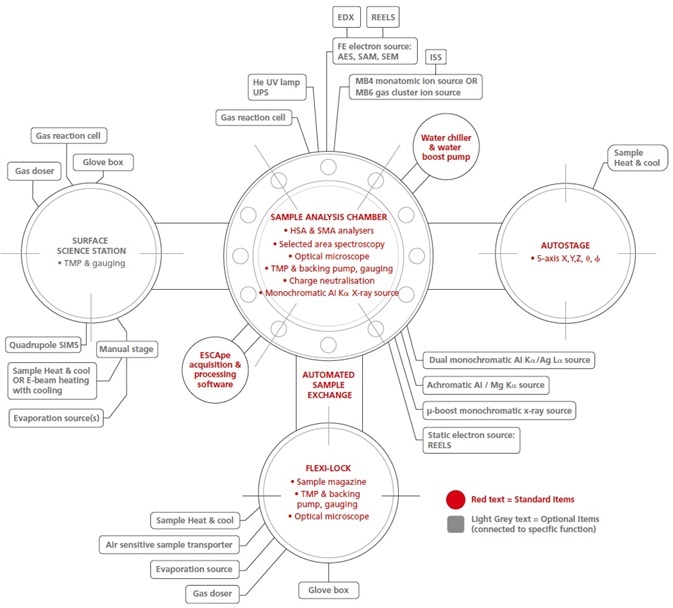
System Configuration Overview
The AXIS Supra+ from Kratos Analytical offers maximum levels of automation along with the flexibility to include complementary sample preparation and/or surface analytical methods. Many optional items illustrated below are now provided as upgrades following the installation.
Kratos Analytical has an established track record in developing surface analysis instrumentation hardware. Mechanical engineers and physicists at Kratos Analytical can offer solutions for surface alteration, sample handling, and further technique incorporation for the AXIS Supra+.
AXIS Supra photoelectron spectrometer