The Amira Software from Thermo Scientific is a robust, comprehensive and adaptable 2D–5D solution specifically developed for imaging, analyzing and comprehending life science and biomedical research data from several image modalities, such as Optical and Electron Microscopy, MRI, CT and other imaging methods.
The Amira Software has unbelievable speed and flexibility, and therefore assists the latest 2D to 5D bioimaging workflows in research areas ranging from structural and cellular biology to tissue imaging, neuroscience, bioengineering and preclinical imaging.
With any 3D image data, such as multi-channel and time series, the Amira Software provides an extensive range of data visualization, processing and analysis capabilities. The Amira Software enables life science and biomedical scientists to gain valuable knowledge about their data, at various scales and from any modality.
Image Credit: Thermo Fisher Scientific - Software
Watch an Introduction to Amira Software
Confidence
The robust segmentation and image processing abilities and workflows, as well as 20+ years of collaboration with the scientific community and thousands of scientists, have currently proven that the digital imaging-based workflows from Thermo Scientific offer trustworthy solutions to biomedical and life science research.
Support
The dedicated professional support team allows users access to the top experts, guaranteeing that no question goes unresolved. Also, with the training, consulting options and ever-expanding collection of tutorials and how-to’s, users can reduce their learning curve and concentrate on getting the answers they require.
Customization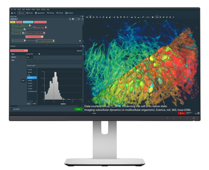
The requirements of users are distinct and keep evolving, therefore the software solutions are completely flexible and open to customization. The scripting interface (Python, TCL), bridge with MATLAB, and the programming API help users to expand their software solution and integrate their own IP (Intellectual Property). Also, if required, the company’s professional service team can help users design unique solutions customized to their requirements.
Automation
The company’s automation abilities and the addition of the continuously expanding online repository of add-ons (Xtra Gallery) help users encapsulate repeatable workflows into simple-to-reproduce recipes.
Thanks to the addition of artificial intelligence, analysis can be executed even by non-image processing experts. It enables them to save time on complex analysis while guaranteeing consistency of results.
Applications
- Cell biology
- Preclinical imaging
- Neuroscience
Watch a Demonstration of Amira Software
Techniques
Correlative imaging
Amira Software has long-standing applications in multi-modality imaging techniques, and is, therefore, the ideal tool for researchers working with data from various imaging modalities. Amira architecture assists multi-volume data processing and visualization from the ground up. The automatic registration tools are combined into the standard edition of Amira Software.
Membrane detection
The Amira Software accelerates segmentation by applying automated detection of cellular features, like membranes and filaments. By carrying out advanced image analysis and statistics on the cryo-tomograms, Amira Software combines data from these individual structures, averaging out noise and increasing contrast, creating a composite model with higher resolution.
Cell detection
Cellular spheroids are 3D cell cultures that represent in vivo tissue better compared to conventional cellular monolayers. This improves the potential of scientists to study diabetes mellitus, cancer, or stem cells among many applications.
But cell analysis in such 3D multicellular objects is not accurately possible, particularly without ruining the object of study.
High content screening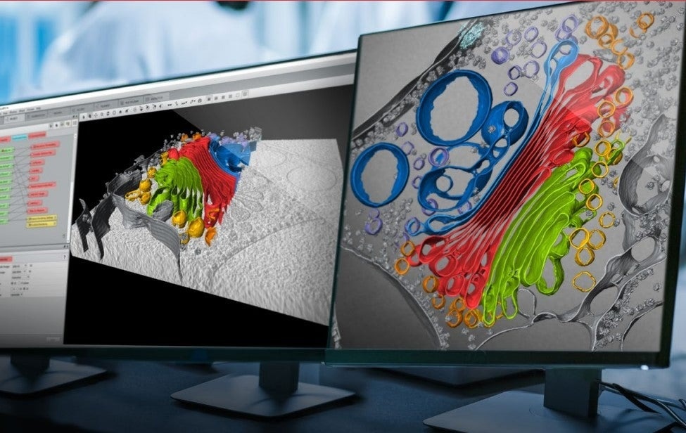
The Amira Software allows multi-channel quantitative capabilities to examine the most demanding scientific and experimental models employed in HCS, such as neuroscience, immuno-oncology, and organoid health. The incredible flexibility and speed of the Amira Software enable the latest 2D–5D bioimaging workflows in various research areas.
Multi-channel and time-series visualization
The latest Xplore5D extension of the Amira Software can be used by life scientists to easily visualize, correlate and process large multi-channel and time-series data in a single platform. This removes concerns regarding image size or memory limits. Thanks to its smart file format, Xplore5D enables users to compress all imaging data without losing valuable information gathered during acquisition.
Filament tracing
The Amira Software helps users to make use of a template-matching algorithm to automatically detect and trace such fine filaments in noisy cryo-EM or DualBeam data. Additionally, the Amira Software allows users to reconstruct filamentous networks and edit the resulting graphs to eliminate image features erroneously determined as filaments or to add missing parts of a network.
3D visualization of a Golgi apparatus from the green alga Chlamydomonas reinhardtii with Amira
3D visualization of a Golgi apparatus from the green alga Chlamydomonas reinhardtii using Amira Software.
Video Credit: Thermo Fisher Scientific - Software
Request a Trial of Amira Software