Beacon Discovery™ enables real-time functional investigation of single cells. Powered by optofluidic technology, it allows researchers to investigate multimodal and temporal cellular responses in real time, easily integrating functional data with sequencing from the same single cell for more profound scientific discoveries.
This user-friendly, small, and adaptable device provides unmatched insights into immune profiling, cell therapy, antibody, and TCR/CAR development.
Why choose Beacon Discovery
Accessible: Lower cost of ownership, lower running costs, and an intuitive interface, enabling easy adoption.
Compact: The benchtop size maximizes laboratory space without losing functionality.
Flexible: Provide comprehensive procedures for immune profiling, antibody development, and customized functional tests.
Proven: Based on an extensively documented and validated optofluidic platform.
Changing the game
Beacon Discovery™ is designed to push the frontiers of live single-cell functional analysis and provides precise control over single-cell experiment design. It permits live cell imaging, several consecutive experiments on the same live cells, and capturing dynamic biological reactions throughout time.
It also recovers high-value cells, allowing researchers to combine on-chip functional insights with off-chip genomes and transcriptomics (TCR-seq, BCR-seq, and mRNA-seq) from the same cells. These features go much beyond what standard platforms can provide.
Applications
Understand the life of single cells
Beacon Discovery™ is a versatile platform for translational researchers in academia and the biopharmaceutical industry to investigate high-parameter, dynamic functional cell analysis. It allows T cell profiling in immunotherapy, antibody identification, and vaccine development.
T cell profiling
Examine T cell activity to identify efficient TCR/CAR sequences, evaluate T cell performance, convert cellular functioning to clinical biomarkers, and recover cells of interest for multi-omic insights.
Assays available: Cytokine analysis, Surface marker staining, Cytotoxicity, Phenotyping
Antibody discovery
Use function-first, high-throughput single B cell screening to transform antibody discovery and increase antibody campaign success.
Assays available: Affinity, cross-reactivity, ligand blocking, antigen specificity, and membrane-bound antigen cell-binding.
Having used the Beacon platform for years, we have seen firsthand how it transforms TCR discovery for cancer immunotherapy. Its automated workflows and novel multi-parameter temporal analysis allow precise characterization of tumor antigen-specific T cells, accelerating the identification of potent therapeutic candidates from patient tumor-infiltrating lymphocytes (TILs). With the new Beacon Discovery platform, this powerful technology is now within reach for many more individual and core labs in academia and industry."
Dr. Joseph Zenga, MD, Associate Professor and Division Chief, Medical College of Wisconsin
Features
Built for easy use and real-time data exploration
Easy-to-use interface
It has never been simpler to design intricate single-cell functional analysis assays. A new, user-friendly wizard-based interface for creating multistep experimental processes is part of the Beacon Discovery™ and is part of the Cell Analysis Software (CAS) Suite. Users can set up and run complex pre-defined and customized workflows with a few clicks.
Deep data analysis
The Cell Analysis Software Suite makes real-time data exploration through automated and manual image analysis possible. Over time, biomolecule secretion, target death, and cell-cell interactions can help researchers make well-informed decisions. Machine learning streamlines cell counts, selection, and segregation. UMAPs simplify multidimensional functional analysis.
Accessible system based on proven optofluidic technology
Key product features
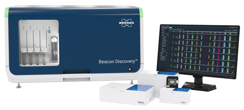
Image Credit: Bruker Cellular Analysis
- Small, benchtop device
- Throughput: 1 chip, up to 20,000 single cells per run
- Four fluorescent channels (exchangeable cubes)
- 4× bright field magnification
- Software that is easy to use and customizable
- All OptoSelect® chips are compatible with it
Technology
Unique features of the beacon technology
The OptoSelect chip, also known as optofluidics, is the central component of Beacon technology. It integrates nanofluidics and Opto-Electro Positioning (OEP) technology. In order to precisely isolate and characterize individual cells at scale, optofluidics uses light and millions of light-actuated pixels to control cells and beads. Afterward, individual cells with desirable characteristics can be extracted for sequencing and other downstream examinations.
Opto-Electrical Positioning (OEP) technology
Provides exquisite control of cells and beads
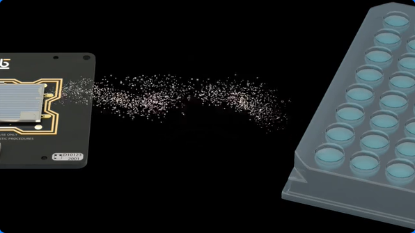
Image Credit: Bruker Cellular Analysis
- Utilizes light to move cells around and into NanoPens
- Allows for thousands of parallel single-cell investigations at once
NanoPen horizontal design
Allows continuous media perfusion
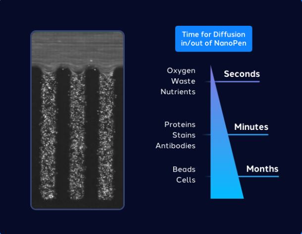
Image Credit: Bruker Cellular Analysis
- Minimizes the disruption of the NanoPens’ contents
- Permits waste, nutrients, and oxygen to be exchanged in and out of the NanoPens in a matter of seconds
- Maintains cells alive and healthy for days or weeks
Flexible to run chips with flexible chip formats
Flexible chip formats allow users to tailor experiments to unique research objectives. The Beacon Discovery™ system is compatible with a variety of chip configurations and adapts effortlessly to a wide range of workflows and applications.
OptoSelect 1750b chip

Image Credit: Bruker Cellular Analysis
This chip contains approximately 1,750 1.7 nL NanoPens suitable for long-term culture experiments, such as adherent cell cloning.
OptoSelect 3500 chip
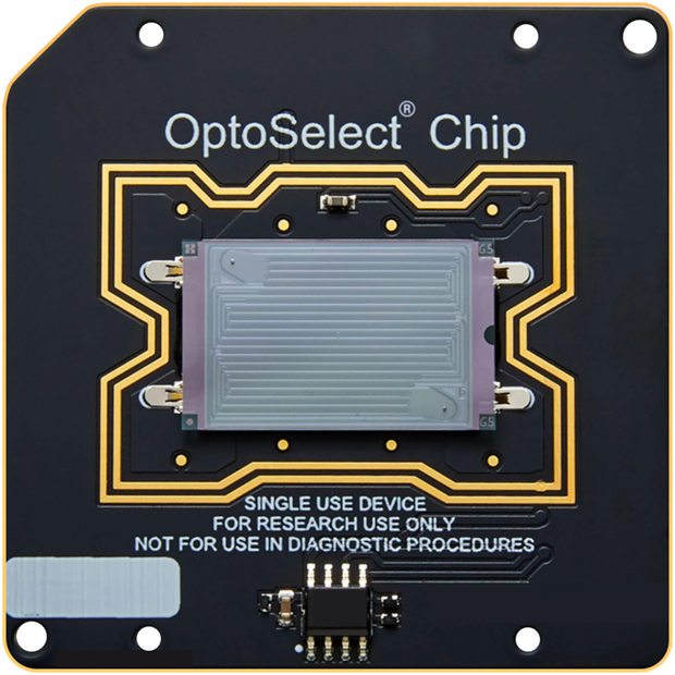
Image Credit: Bruker Cellular Analysis
This device has around 3,500 chambers with 0.75 nL NanoPens, allowing cell division and increased clone throughput.
OptoSelect 11k chip
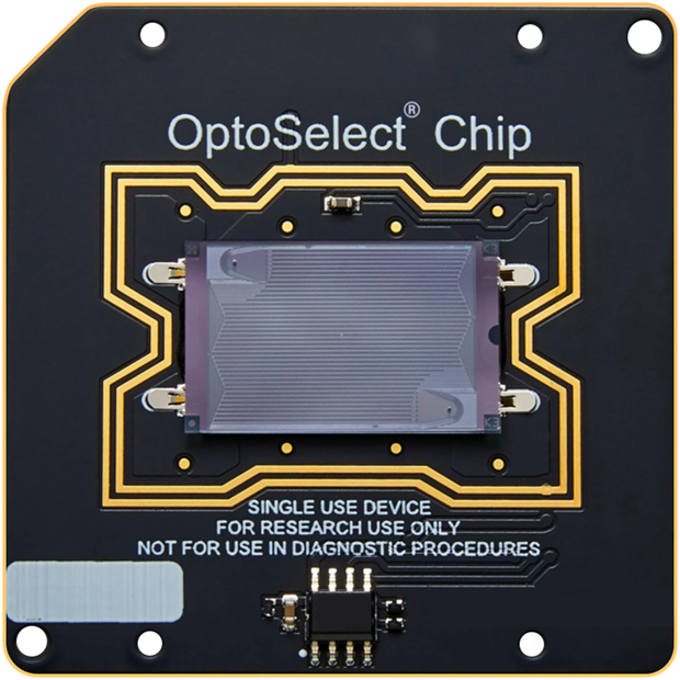
Image Credit: Bruker Cellular Analysis
This device has around 11,000 chambers with 0.32 nL NanoPens, allowing for increased throughput in single-cell screening.
OptoSelect 20k chip
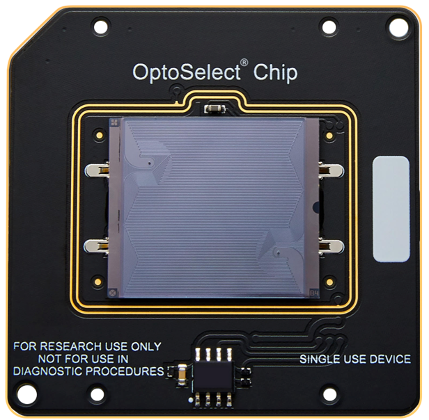
Image Credit: Bruker Cellular Analysis
This chip comprises approximately 20,000 chambers with 0.32 nL NanoPens for high-throughput single-cell screening.
How it works
Streamlined workflow for rapid discoveries
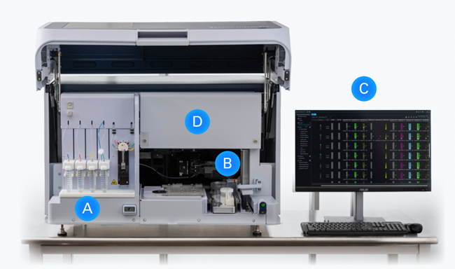
Image Credit: Bruker Cellular Analysis
The system combines configurable Bruker Cellular Analysis reagents with OptoSelect chips to provide a consistent user experience.
Load Reagents and Samples - Put the reagents in the reagent bay (A) and OptoSelect chips in the sample bay (B).
Guided Workflow Setup - The Workflow Wizard (C) guides users through the experiment setup and execution.
Exquisite Control on Single-Cell Experiments - OEP technique softly moves and arranges live single cells and reagent beads in NanoPens, resulting in precise functional characterization.
Multi-Modal and Temporal Analysis with Live Single-Cell Imaging - The OptoSelect chip and horizontal NanoPen design allows continuous perfusion and quick reagent exchange, allowing cells to live for days or weeks. Run numerous experiments on the same cells at various time points. Four exchangeable fluorescent channels (D) provide for real-time brightfield and fluorescence imaging with minimal hands-on time.
Data Capture and Sequencing Integration - Identify and recover high-value cells, then apply functional insights to transcriptomic and genomic sequencing for further investigation.