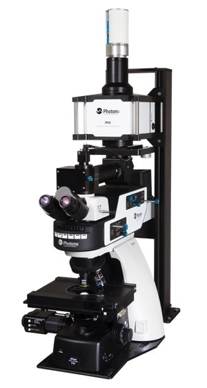
The IMA™ from Photon etc. is a hyperspectral microscope that offers spectral and spatial data in the VIS, NIR and SWIR ranges (from 400 to 1620 nm). This technology maps fluorescence, reflectance and transmittance very fast. IMA is quicker and more effective than typical point-by-point or line-scan-based systems thanks to its high-throughput global-imaging filters. It is useful for biological analysis and research on organic and inorganic substances.
Key features
- Quick global mapping (non-scanning)
- Absolute photometric calibration
- Excellent spatial and spectral resolution
- Non-destructive testing under real-world conditions
- Extremely adaptable
- Complete system (camera, illumination module, filters, microscope and software)
Photon etc - IMA Hyperspectral Microscope
IMA Global Hyperspectral Microscope: Video Credit: Photon etc.
Applications overview
- IR markers in complex environments such as live cells and tissue can be studied, for instance, the spectral heterogeneity of IR fluorophores produced in the second biological window
- Obtaining darkfield images and contrast of transparent and unstained samples like living cells, polymers or crystals
- Multicolor markers tracking (deep multiplexing in vivo)
- Recognizing disease-affected tissues
- Cellular environment monitoring
Specific applications
- Optical biosensor for detecting Escherichia coli concentrations
- Using single-walled carbon nanotubes (SWCNT) as fluorescent probes for multiplexed imaging
- Tracking of various labelled receptor subtypes in live neurons
- Specifying isolated carbon nanotube species' molar concentrations
- Mapping of endolysosomal lipid flux
- Reporting of an enzymatic suicide inactivation pathway
- Assessment of nitric oxide concentration
- Identification and localization of gold nanoparticles (AUNP) using hyperspectral darkfield characterization
- Reporting of microRNA hybridization events in vivo
- Nuclear entry reporting via a noncanonical pathway
- Identification of colon cancer metastasis in the liver whilst operating
- Biomolecular functionalization to control live-cell stability and retention
- Drug screening for CNS diseases by screening multiple protein subtypes simultaneously and live tracking of their spatial dynamics
- Investigating the permeation of multicellular tumor spheroids
- Ascertaining the molar concentrations of isolated carbon nanotube species
- Monitoring of different receptor subtypes labelled in live neurons
- Accelerated detection of Escherichia coli in phosphate-buffered saline solution
Product specifications
Source: Photon etc.
| |
VIS |
SWIR |
| Spectral range |
|
|
| Spectral resolution (FWHM) |
|
|
| Spectral channels |
|
|
| Spatial resolution |
- Sub-micron - limited by the microscope objective NA
|
- Sub-micron - limited by the microscope objective NA
|
| Camera |
|
- Photon etc. InGaAs camera (ZephIR™ 1.7 or Alizé™ 1.7)
|
| Excitation wavelengths (up to 3 lasers) |
- 405, 447, 532, 561, 660, 730, 785, 808 nm (other wavelengths available upon request)
|
- 405, 447, 532, 561, 660, 730, 785, 808 nm (other wavelengths available upon request)
|
| Microscope |
- Upright or inverted, scientific grade
|
- Upright or inverted, scientific grade
|
| White light illumination |
- Diascopic, episcopic, Hg, halogen
|
- Diascopic, episcopic, Hg, halogen
|
| Illumination options |
- Epifluorescence module, darkfield module (oil or dry)
|
- Epifluorescence module, darkfield module (oil or dry)
|

Image Credit: Photon etc.