In a recent webinar, Dr. Lena Smirnova explained how brain microphysiological systems are transforming in vitro neurotoxicity testing and disease modeling. NewsMedical compiled the key highlights in the interview below.
Can you please introduce yourself and your role at Johns Hopkins University?
I'm Lena Smirnova, Assistant Professor at the Department of Environmental Health and Engineering, Bloomberg School of Public Health at Johns Hopkins University.
My work is centered around developing human-relevant models, with a specific focus on brain microphysiological systems, to improve chemical safety and drug testing, particularly in the context of developmental neurotoxicity and disease modeling.
What are brain microphysiological systems (bMPS), and why are they so important in toxicology and neuroscience research?
bMPS are 3D models, such as organoids, assembloids, and organ-on-chip platforms, derived from human induced pluripotent stem cells. They aim to replicate the architecture and function of the human brain, capturing key features like network formation and cellular diversity.
These models overcome some limitations of 2D cultures and animal models, and so provide more physiologically relevant insights into human neurodevelopment and disease.
What key limitations of animal models do bMPS help overcome?
Animal models are expensive, time-consuming, and often fail to replicate human-specific biological processes.
For instance, a single Developmental Neurotoxicity (DNT) study in animals can cost over a million dollars and use more than a thousand rats. bMPS enable higher-throughput testing and allow for studying human-relevant responses, including those linked to sex, genetics, and immune function. They are also better suited for long-term and low-dose exposure studies.
How are you using high-content imaging and gene editing to assess neurodevelopment in bMPS?
We employ CRISPR/Cas9 gene editing to generate fluorescent reporter lines for tracking specific cell populations or developmental events in real-time. High-content imaging then allows us to measure a variety of markers, such as proliferation, apoptosis, and synaptogenesis, across a developmental timeline.
This approach supports efficient and reproducible assessment within the same 3D model.
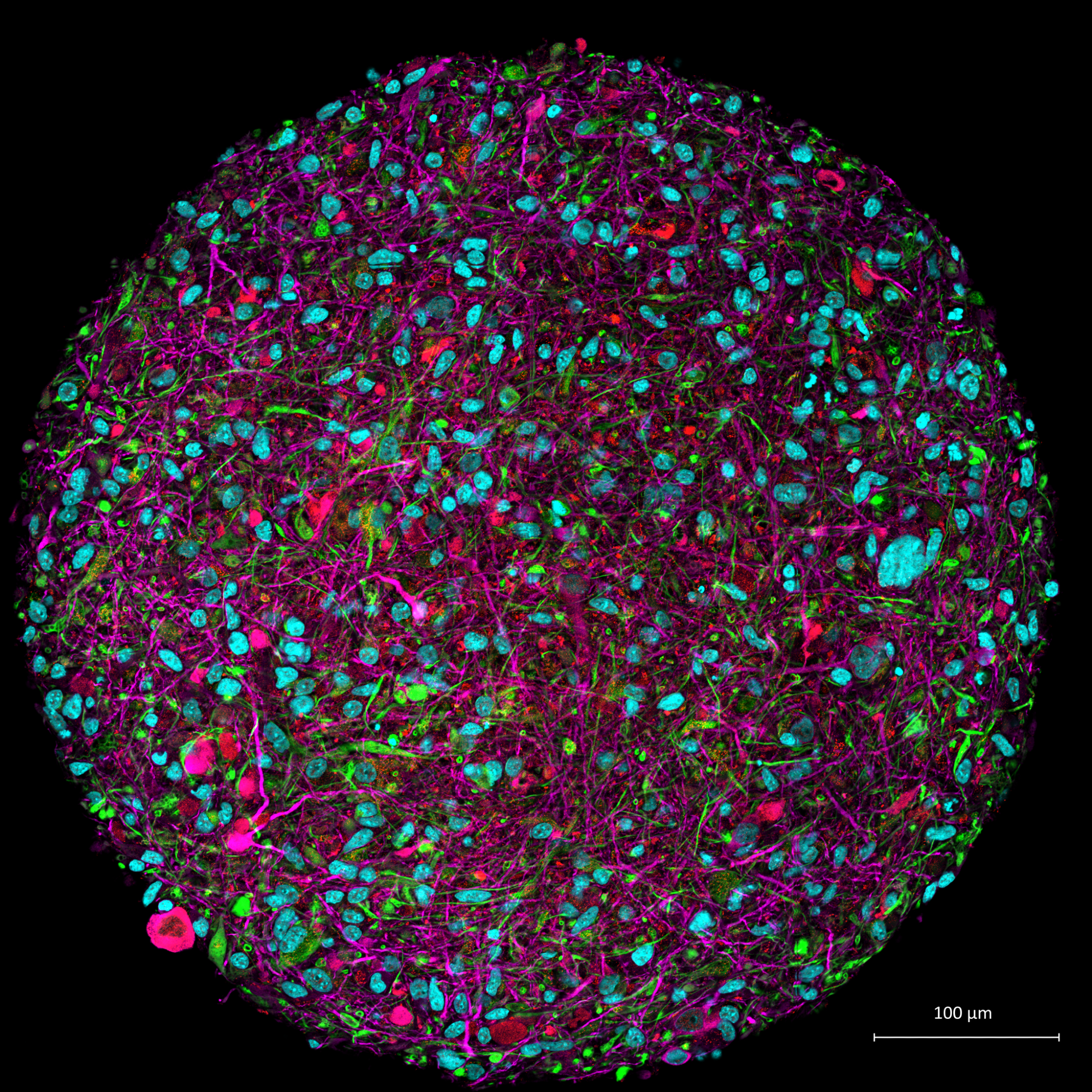
Image Credit: Thomas Hartung, Johns Hopkins University
What are some recent case studies where bMPS have provided new insights into neurotoxicity?
In one study, we examined the effects of cadmium and chromium, both found in drinking water, and observed disrupted viability and neurite outgrowth.
Another case involved chronic exposure to low doses of domoic acid, which led to impaired network synchrony and plasticity. These effects were detectable even at doses that were not overtly cytotoxic, highlighting subtle but critical neurotoxic impacts.
How do you model functional brain activity, like learning and memory, in vitro?
We've developed an "organic intelligence" concept that combines bMPS with high-density microelectrode arrays and machine learning. This allows us to design open-loop and closed-loop experiments where brain organoids can be trained to recognize patterns or interact with simulated environments. With this type of assay, we are aiming to develop in vitro analog for cognitive functions typically assessed in animal behavior tests.
Can bMPS be used for personalized or sex-specific neuroscience research?
Yes. Because they are derived from donor-specific iPSCs, bMPS can reflect sex-based biological differences or genetic variants associated with disease.
We've also integrated immune cells like microglia into our models, enabling us to explore neuroinflammatory mechanisms relevant to autism and other conditions. This opens up possibilities for precision medicine and targeted toxicology.
What are some challenges that still need to be addressed in bMPS development?
Challenges include the need for skilled handling, time-intensive differentiation protocols, and iPSC variability. There's also a pressing need to adapt traditional 2D assay endpoints for use in 3D contexts. Standardization across labs is essential to improve reproducibility and build confidence in these models for regulatory purposes.
How do you see brain microphysiological systems integrating into regulatory toxicology frameworks?
A growing interest is emerging around integrating bMPS into regulatory science. The OECD has already adopted in vitro methods for DNT testing, and this work builds on that foundation by incorporating more complex endpoints.
High content imaging is used extensively, with the Yokogawa CQ1 combined with CRISPR-Cas9 to generate reporter cell lines that enable faster, more reproducible, and reliable assessment of key neural development events.
The goal is to move toward harmonized, validated platforms that deliver high-confidence data for chemical safety decisions while reducing reliance on animal studies.
What message would you share with researchers considering bMPS for their work?
Invest in quality. These models are complex, but complexity can't make up for poor cell quality or inconsistent protocols. Make sure you have the right tools and quality controls in place. Done right, bMPS offer a powerful way to explore human brain development and neurotoxicity in a much more relevant context than traditional models.
About Dr. Lena Smirnova
Dr. Lena Smirnova is an Assistant Professor at the Bloomberg School of Public Health, Johns Hopkins University, in the Department of Environmental Health and Engineering. She earned her PhD from Charité University in Berlin, Germany, and completed her postdoctoral training at the Federal Institute for Risk Assessment in Berlin, Germany.
Dr. Smirnova is a founding member and past-president of the International Microphysiological Systems Society and is currently a strong advocate for the advancement and regulatory acceptance of non-animal methods in toxicology. Her interdisciplinary approach bridges neuroscience, stem cell biology, and toxicology to develop next-generation tools for brain research and chemical safety assessment.
About Yokogawa Life Science Solutions

Yokogawa provides advanced imaging and automation technologies to accelerate research and drug discovery. Our high-content imaging systems and cell analysis platforms deliver precise, high-throughput data for deeper insights into cellular processes - empowering breakthroughs in your research. .
 Yokogawa introduces CellVoyager High-Content Analysis System CQ3000