Prototype systems used in developmental biology use a wide range of transgenic animals, in which individual cells, specific tissues or whole embryos are labeled with fluorescent proteins. These fluorescent transgenic organisms make it possible to observe the behavior of cells and tissues during developmental processes, in real time and at high resolution. Such observations may lead to a better understanding about the various mechanisms involved in shaping a complex organism. This is something that is often limited by the technical limitations of imaging apparatus.
In-vivo imaging has the potential to obtain quantitative information at a single-cell resolution. When this imaging technique is carried out non-invasively on fully functioning and intact organisms, time lapse microscopy enables developmental processes to be studied over time.
Fluorescence Microscopy
The speed, resolution and penetration of fluorescence microscopy techniques have become increasingly powerful, although only when applied to thin and transparent samples. It is particularly difficult to achieve single cell resolution when examining whole embryos, which are usually a few millimeters in size. Moreover, the resolution achieved when dealing with live specimens is often lower when compared to fixed samples due to the scattering of opaque and intact tissue, the sample size, the movement of organs, pigmentation in untreated animals, and the physiologically sustainable conditions the sample needs to be maintained at. These constraints mean researchers must fix and section the samples, despite the fact that many dynamic developmental processes can only be fully observed in the intact living embryo.
Selective Plane Illumination Microscopy
Selective plane illumination microscopy (SPIM) is a fluorescence microscopy method in which a focused light-sheet is used to light up the sample from the side. Using this technique, a high resolution at high penetration depths can be achieved, while also only being minimally invasive. SPIM provides several benefits when compared to other established methods and these include a high acquisition speed, a high dynamic range, and significantly reduced photo-bleaching. These advantages make SPIM a popular technique for the study of development. Figure 1 shows the schematics of SPIM with two detection and illumination directions.
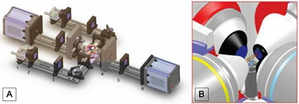
Figure 1. Schematics of selective-plane illumination microscope with two illumination and detection directions.
The concept behind SPIM and other light-sheet-based microscopy methods is to light up the specimen from the side in a well-defined volume around the detection optics’ focal plane. Although different implementations of this concept exist, the overall principles remain the same.
In the SPIM technique, a sheet of varying thickness is produced using cylindrical optics. The light converges towards and diverges away from the sample. The waist of the light sheet is placed in the middle of the field of view (FOV) and the size of the light sheet can be adjusted to suit various different sample sizes. For samples measuring 20 to 100 µm, the sheet can be made as thin as ~ 1 um, while for samples measuring 1 to 5 mm, the sheet can be made thicker, at around 5 to 10 µm, to ensure it remains relatively uniform across the FOV.
A major benefit in SPIM is that only the part of the sample that is in-focus gets exposed to the laser light, which allows for optical sectioning and significantly reduced photodamage. In addition, the fluorescence signal generated by the in-focus area is picked up in parallel for the entire FOV, therefore providing fast imaging speeds.
In light-sheet microscopes, the optical penetration depth is restricted by light scattering. Therefore, simultaneous multiview light sheet microscopy has been developed to overcome this problem. This method removes the spatiotemporal artifacts that occur due to slow sequential multiview imaging as well as introducing new and powerful capabilities.
Simultaneous multiview SPIM can quickly image large fluorescent samples from various directions across biologically relevant timescales using Andor’s sCMOS technology. This counteracts the limitations of sequential multiview strategies and allows quantitative systems-level imaging of the rapid dynamic processes that occur in large living samples.
Andor Solutions for SPIM
Andor’s complete range of sCMOS cameras serve as perfect detectors for SPIM. Both the Zyla 5.5 and Neo 5.5 scientific CMOS (sCMOS) cameras have a 5.5 megapixel sensor with a small pixel size of 6.5 µm that provides high resolution and a large FOV without affecting the frame rate or read noise. The Zyla 4.2 features a 4.2-megapixel sensor with a QE of 72 % at 600 nm. LightScan PLUS (LSP) with flexiScan and CycleMax is a new feature on Zyla 4.2 which is ideal for SPIM. LSP reduce’s background and improve’s contrast and resolution in scattering samples. It is designed to allow users to optimise signal strength and confocality concurrently in applications such as Scanned Light Sheet Microscopy and Line Scanning Confocal Microscopy..,
Conclusion
sCMOS is an advanced technology that provides excellent performance features that make it ideal for quantitative and high fidelity scientific measurement. Additionally, the 5.5 megapixel sensor provides high resolution and a large FOV without affecting frame rate, read noise, or dynamic range. Rolling and Global shutter readout allows for maximum application flexibility. In addition, the Andor Software Development Kit (SDK) allows programmers access to the Andor camera range. Importantly, the SDK has a dynamic link library that can be utilized with a range of programming environments such as LabVIEW, MATLAB, Visual Basic, C, C++ and C#.
About Andor Technology
 Andor Technology, an Oxford Instruments company, is a global leader in the pioneering and manufacturing of high performance scientific imaging cameras, spectroscopy solutions and microscopy systems for research and OEM markets. Andor has been innovating the photonics industry for over 20 years and continues to set the standard for high performance light measuring solutions, enabling its customers to break new ground by performing light measurements previously considered impossible. Andor’s digital cameras, are allowing scientists around the world to measure light down to a single photon and capture events occurring within 1 billionth of a second.
Andor now has over 400 staff across 16 offices worldwide, distributing products to over 10,000 customers in 55 countries. Andor’s products are used in a wide range of applications including medical research to further the understanding of heart disease, cancer and neuronal diseases such as Alzheimer’s and Parkinson’s disease. Andor also has applications for forensic science and astronomy. Through continuous dialogue with customers and strong teamwork, Andor continues to innovate ground-breaking products that improve the world in which we live.
Sponsored Content Policy: News-Medical.net publishes articles and related content that may be derived from sources where we have existing commercial relationships, provided such content adds value to the core editorial ethos of News-Medical.Net which is to educate and inform site visitors interested in medical research, science, medical devices and treatments.