Thermo ScientificTM Amira-AvizoTM Software is an advanced, influential 3D segmentation and visualization software package. Fast processing of complex data sets and an extensive set of 3D modeling tools make it well-suited to various applications in paleontological and biological research.
Dr. Laura Porro explains how Amira-Avizo Software is crucial to her research in integrative anatomy: “I have consistently found Amira-Avizo Software to be the best-suited visualization software for my research needs.”
Computed tomography (CT) is a key technique in anatomy, facilitating non-destructive imaging of biological structures as well as identifying the density of different tissues.
For over a decade, Dr. Laura Porro of University College London has been putting Amira-Avizo Software to use in order to process a wide variety of anatomical CT data.
“I use Amira-Avizo Software to process medical imaging data of humans and living animals to obtain information on skeletal shape and create 3D models,” says Dr. Porro.
“More recently, my colleagues and I have been using various stains and contrast-enhanced CT scanning along with Amira-Avizo Software to visualize soft tissue anatomy in a range of different species, from fish and frogs to reptiles and birds.”
Utilizing chemical stains in combination with micro-computed tomography (µCT) allows for the ‘digital dissection’ of specimens: thus, comprehensive 3D reconstructions of soft- and hard-tissue anatomy can be extracted and analyzed without the necessity of having to ever pick up a scalpel.
In addition to being non-destructive, these techniques allow the visualization of extremely small and fragile specimens without changing the 3D topology of anatomical structures.
Images published in Porro’s include the digital dissections of two teleost fish, the head of the rock dove (Columba livia) and the African clawed frog (Xenopus laevis).1–3
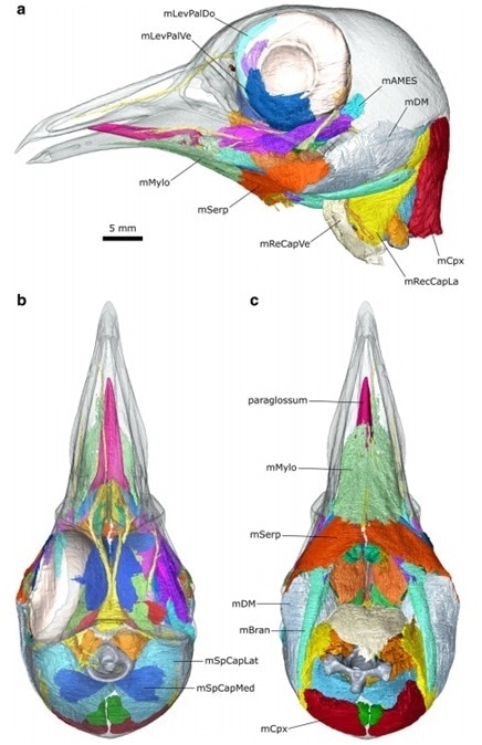
Digital dissection of the head of Columba livia. A) lateral view, B) dorsal view, C) ventral view. Ref: Jones, M.E.H., Button, D.J., Barrett, P.M. et al. Digital dissection of the head of the rock dove (Columba livia) using contrast-enhanced computed tomography. Zoological Lett 5, 17 (2019).1
“Our greatest challenge is posed by segmenting imaging data of fossil animals, which often features poor contrast between fossil bones and surrounding rock,” states Porro.
“Over the years, Amira-Avizo Software has repeatedly proven to be the best solution for processing these difficult data sets, providing anatomical data that is fundamental for understanding the shape of the tree of life.”
Amira-Avizo Software can be utilized to process µCT data and generate detailed interactive 3D reconstructions for large-scale distribution to the public, students, and researchers.
A suite of 3D image processing tools within Amira-Avizo Software can be used to recast CT data from fossil samples, facilitating reconstructive manipulation of 3D models.
Porro and her colleagues make use of these tools to ‘digitally restore’ fossils of extinct species, allowing them to “see for the first time how these remarkable animals appeared in life.”4,5
Porro further states, “The interpolation, smoothing and transformation tools available within Amira-Avizo Software are ideal for manipulating elements in 3D space, allowing us to remove millions of years of damage and deformation…”
Using Amira-Avizo Software, Porro and her team were able to identify the bone structures of certain species using detailed segmented scans of fossil specimens.
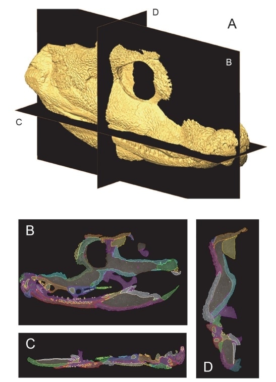
Segmentation of CT data of the extinct tetrapod Acanthostega gunnari. Ref: Porro LB, Rayfield EJ, Clack JA (2015) Descriptive Anatomy and Three-Dimensional Reconstruction of the Skull of the Early Tetrapod Acanthostega gunnari Jarvik, 1952. PLoS ONE 10(3): e0118882.4
“Tools within Amira-Avizo Software are essential for preparing and exporting models for further applications including 3D morphometric analyses and biomechanical modelling.”
The 3D models generated by the Amira-Avizo Software are not just practical for visualization. Porro and her colleagues employed the software to reconstruct the musculoskeletal anatomy of the skull of the extinct marine crocodile Pelagosaurus for a comparison with the skull of the living Indian gharial Gavialis.
Finite element analysis of the resulting 3D model facilitated the investigation of stress distribution in the skull, providing insight into the hunting and eating habits of Pelagosaurus.6
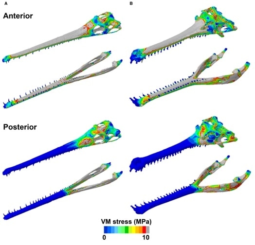
Von Mises stress distribution plots of the skulls of Pelagosaurus (A) and Gavialis (B), simulating a unilateral biting scenario at the anterior and posterior tooth positions. Grey areas represent von Mises stress values higher than 10 MPa. Ref: Ballell, A., Moon, B.C., Porro, L.B., Benton, M.J. and Rayfield, E.J. (2019), Convergence and functional evolution of longirostry in crocodylomorphs. Palaeontology, 62: 867-887.6
Utilizing 3D models generated by Amira-Avizo Software for comparison of finite element modeling of the mandible of Alligator mississippiensis demonstrated that this approach closely matches experimental in vivo results.7
Porro used Amira-Avizo Software to repair the skeleton of an extinct animal specimen at UCL’s Grant Museum of Zoology in a recent project, calling attention to the intrinsic value of straightforward and precise 3D modeling for education and collaboration.
The specimen on the agenda – the skeleton of a quagga Equus quagga, a relative of the living zebra – had its left hind leg missing. By scanning and mirroring the existing right hind leg, a 3D model was created from which a 3D-printed replacement could be generated.8
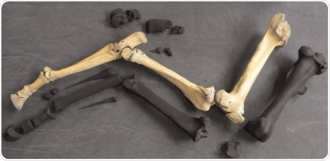
The rear right limb of an Equus quagga quagga skeleton along with the 3D printed models of individual mirrored bones ready for assembling. Ref: Nigel R. Larkin, Laura B. Porro; Three legs good, four legs better: Making a quagga whole again with 3D printing. Collection Forum 1 January 2016; 30 (1-2): 73–84.8
Amira-Avizo Software offers a comprehensive solution for each step of the workflow, from segmenting of complex and intricate images to 3D rendering and physical modeling.
Dr. Porro concludes, “Amira-Avizo Software is an absolutely essential tool for carrying out cutting-edge anatomical and biomechanical work within our research group, one I anticipate we will continue using for many years to come.”
To discover more about the features, Amira-Avizo Software offers or to book a free trial, contact the Thermo Fisher team today.
References
- Jones, M.E.H., Button, D.J., Barrett, P.M. et al. Digital dissection of the head of the rock dove (Columba livia) using contrast-enhanced computed tomography. Zoological Lett 5, 17 (2019). https://doi.org/10.1186/s40851-019-0129-z.
- Brocklehurst R, Porro L, Herrel A, Adriaens D, Rayfield E. A digital dissection of two teleost fishes: comparative functional anatomy of the cranial musculoskeletal system in pike (Esox lucius) and eel (Anguilla anguilla). J Anat. 2019 Aug;235(2):189-204. doi: 10.1111/joa.13007.
- Porro LB, Richards CT. Digital dissection of the model organism Xenopus laevis using contrast-enhanced computed tomography. J Anat. 2017 Aug;231(2):169-191. doi: 10.1111/joa.12625.
- Porro LB, Rayfield EJ, Clack JA (2015) Descriptive Anatomy and Three-Dimensional Reconstruction of the Skull of the Early Tetrapod Acanthostega gunnari Jarvik, 1952. PLoS ONE 10(3): e0118882. https://doi.org/10.1371/journal.pone.0118882
- Porro LB, Witmer LM, Barrett PM. Digital preparation and osteology of the skull of Lesothosaurus diagnosticus (Ornithischia: Dinosauria). PeerJ. 2015 Dec 21;3:e1494. doi: 10.7717/peerj.1494.
- Ballell, A., Moon, B.C., Porro, L.B., Benton, M.J. and Rayfield, E.J. (2019), Convergence and functional evolution of longirostry in crocodylomorphs. Palaeontology, 62: 867-887. https://doi.org/10.1111/pala.12432.
- Porro LB, Metzger KA, Iriarte-Diaz J, Ross CF. In vivo bone strain and finite element modeling of the mandible of Alligator mississippiensis. J Anat. 2013;223(3):195-227. doi:10.1111/joa.12080.
- Nigel R. Larkin, Laura B. Porro; Three legs good, four legs better: Making a quagga whole again with 3D printing. Collection Forum 1 January 2016; 30 (1-2): 73–84. doi: https://doi.org/10.14351/0831-4985-30.1.73.
About Thermo Fisher Scientific – Electron Microscopy Solutions
 With more than 60 years of innovation and leadership, FEI enables customers to find meaningful answers to questions that accelerate breakthrough discoveries, increase productivity, and ultimately change the world. FEI designs, manufactures, and supports the broadest range of high-performance microscopy workflows that provide images and answers in the micro-, nano-, and picometer scales.
Combining hardware and software expertise in electron, ion, and correlative microscopy with deep application knowledge in the materials science, life sciences, electronics, and natural resources markets, the worldwide FEI team of 2,700+ employees is dedicated to customers’ pursuit of discovery and resolution to global challenges.
Thermo Fisher Scientific Electron Microscopy Overview | From Materials to Life Sciences
Sponsored Content Policy: News-Medical.net publishes articles and related content that may be derived from sources where we have existing commercial relationships, provided such content adds value to the core editorial ethos of News-Medical.Net which is to educate and inform site visitors interested in medical research, science, medical devices and treatments.