Photoacoustic microscopy (PAM) is an advanced in vivo tissue imaging technique that combines optical and acoustic approaches to overcome the optical diffusion limit.
It can produce images with great spatial resolution at depths of up to several millimeters and can simultaneously image multiple contrasts. For example, these approaches and contrasts could be used for anatomical, functional, flow dynamic, metabolic, and molecular picture modalities.
How does photoacoustic microscopy work?
Photoacoustic microscopy starts with light - usually a nanosecond pulsed laser beam. This brief pulse of energy initiates the photoacoustic effect: photons absorbed by tissue generate a slight, localized temperature rise. As the tissue undergoes thermoelastic expansion, it emits a broadband acoustic wave, which can be captured using ultrasound detection.
The acoustic energy is then converted into a voltage signal, producing a one-dimensional, depth-resolved image known as an A-line for each laser pulse. By performing 2D raster scanning across the sample, multiple A-lines are combined to construct a full 3D photoacoustic image.
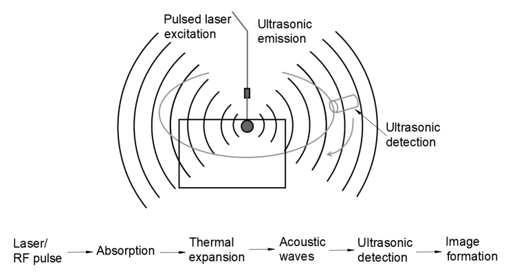
Photoacoustic microscopy uses both light and acoustic methods to produce an image more detailed than could be produced by either method alone. Image Credit: Avantier Inc.
The key to in vivo imaging with PAM lies in two fundamental properties: the deep penetration of diffused photons and the extremely low scattering of sound, about 1,000 times less than that of light.
Together, these allow researchers to achieve imaging depths that would be impossible with optical methods alone, while also providing precise control over penetration depth. This versatility makes it possible to use the same technique for high-resolution imaging of both a mouse ear and a mouse brain. In PAM, axial resolution is determined by the bandwidth of the ultrasound transducer.
Photoacoustic microscopy can be divided into two main types. Optical-resolution PAM (OR-PAM) is used when the optical focus is tighter than the acoustic focus, making it ideal for imaging up to ~1 mm deep. Acoustic-resolution PAM (AR-PAM), where the acoustic focus dominates, is better suited for depths of 1–3 mm.
The contrast of PAM images is dictated by the optical properties of the tissue. For non-invasive imaging, intrinsic absorbers such as red blood cells, DNA, lipids, and glucose provide natural contrast. For other applications, researchers can introduce exogenous agents - such as organic dyes, nanoparticles, or fluorescent proteins - to enhance visualization.
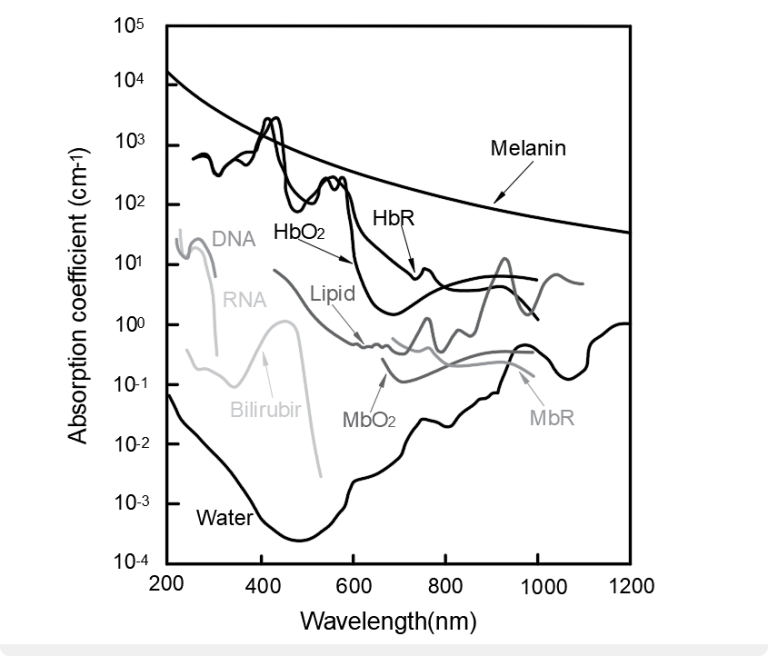
Photoacoustic microscopy can produce high resolution images with endogenous contrast agents. This graph compares the absorption spectra of some of the more common contrast agents found in biological tissue. Image Credit: Avantier Inc.
Applications of photoacoustic microscopy
PAM imaging plays a vital role in both medicine and research, offering a way to see inside tissues that would otherwise appear opaque.
In clinical contexts, it can aid diagnostics by providing detailed information on blood flow, oxygen metabolism, or tumor progression. It also has surgical applications, where it can guide precise incisions, as well as research applications, where it helps uncover the structural and functional changes associated with disease.
A clear example of PAM’s value in medical research is its use in studying inflammatory skin diseases. When combined with optical coherence tomography, PAM enabled researchers to identify oxygenation differences, thickening of the epidermis, and disorganized vascular patterns with dilated vessels, along with the absence of melanin in eczematous skin tissue.
Photoacoustic microscopy at Avantier
Avantier specializes in manufacturing high-quality custom optics for applications such as photoacoustic microscopy. Its clients have been able to push the limits of existing technology, resulting in innovative imaging speeds and quality.
For example, in a recent Science Advances paper titled Optical-Resolution Parallel Ultraviolet Photoacoustic Microscopy for Slide-Free Histology, the authors used a custom F-theta lens from Avantier to reach the imaging speeds necessary for potential real-time intraoperative photoacoustic histology.
Do you want to take your optical project to the next level? Avantier’s expert engineering and design teams are ready to collaborate with you to design and manufacture the exact optical components or systems your application demands.
References
- Attia, A.B.E., et al. (2019). A review of clinical photoacoustic imaging: Current and future trends. Photoacoustics, 16, p.100144. https://doi.org/10.1016/j.pacs.2019.100144.
- Ning, B., et al. (2015). Ultrasound-aided Multi-parametric Photoacoustic Microscopy of the Mouse Brain. Scientific Reports, 5(1). https://doi.org/10.1038/srep18775. .
- Cao, R., et al. (2024). Optical-resolution parallel ultraviolet photoacoustic microscopy for slide-free histology. Science Advances, 10(50). https://doi.org/10.1126/sciadv.ado0518. .
- Yao, J. and Wang, L.V. (2013). Photoacoustic microscopy. Laser & Photonics Reviews, 7(5), pp.758–778. https://doi.org/10.1002/lpor.201200060.
- Zabihian, B., et al. (2015). In vivo dual-modality photoacoustic and optical coherence tomography imaging of human dermatological pathologies. Biomedical Optics Express, 6(9), p.3163. https://doi.org/10.1364/boe.6.003163. .
About Avantier Inc
Avantier is a vertically integrated leader in high-precision custom optics, delivering end-to-end solutions for complex optical challenges. From prototyping to full-scale production, we ensure exceptional quality and repeatability.
Our expertise spans the design and manufacturing of advanced optical components such as aspherical lenses, freeform optics, microscope objective lenses, OAP mirrors, telescopes, SiC optics, optical domes, microlens arrays, large optics and infrared lenses.
Serving aerospace, biomedical, life sciences, quantum computing, and other advanced technology sectors, Avantier is your trusted one-stop strategic partner for meeting the most stringent optical specifications with precision and reliability.
Sponsored Content Policy: News-Medical.net publishes articles and related content that may be derived from sources where we have existing commercial relationships, provided such content adds value to the core editorial ethos of News-Medical.Net which is to educate and inform site visitors interested in medical research, science, medical devices and treatments.