Key takeaways
- Medical lens-based ear endoscopes enable high-resolution visualization, enhancing the diagnosis and treatment of ear diseases.
- These devices use sophisticated optical and illumination systems for in-depth examination and surgical support.
- Ongoing developments, including 3D imaging and AI integration, promise further improvements in this essential otolaryngology instrument, enhancing diagnostic precision and surgical outcomes.
Advances in medical technology have established endoscopy as a pivotal instrument for minimally invasive examination of internal anatomical structures, providing both diagnostic and therapeutic benefits.
Unlike other imaging techniques, it offers direct visualization, allowing clinicians to detect even the subtlest abnormalities. More recently, rigid and versatile lens-based ear endoscopy has become central to the diagnodiagnosing and treatingdisorders.
Fundamental principles of lens-based ear endoscopy
Lens-based ear endoscopes are specialized optical tools engineered for in-depth assessment of the external auditory canal (EAC), tympanic membrane (TM), and middle ear. These regions have a complicated anatomical structure. The ossicular chain, for example, ofpreventsy of visualization, requiring sophisticated imaging methodsobtain get a better view.
The introduction of high-resolution, small-diameter endoscopes has transformed otolaryngology by providing direct, magnified views of these structures, supporting improved diagnostic precision, and enabling targeted therapeutic interventions.
Operational mechanisms of lens-based ear endoscopes
1. Optical imaging principles:
Contemporary ear endoscopes use compact, high-definition camera systems integrated at the distal tip. These devices often include multi-element lens stacks designed to optimize resolution, depth of field, and field of view within the confined dimensions of the ear canal. Integrated light sources provide illumination, while the image sensor captures reflected light from the target tissue (e.g., CMOS or CCD).
Analog signals are then processed and converted into digital video for instant display on a monitor.. Sophisticated systems typically use digital image processing algorithms to improve image clarity, color fidelity, and contrast to aid in differentiating healthy and pathological tissues.
2. Illumination systems:
High-quality visualization in the narrow and often obstructed ear canal relies on effective illumination. Modern ear endoscopes largely use cold light sources, such as high-intensity LEDs, paired with fiber optic light guides for safe and efficient light delivery to the distal tip. This design reduces thermal output at the tissue interface, minimizing the risk of iatrogenic thermal damage to fragile otologic structures.
Innovative illumination techniques, such as narrow-band imaging (NBI), are transforming endoscopy. By exploiting light absorption differences, they improve the detection of vascular patterns and mucosal abnormalities.
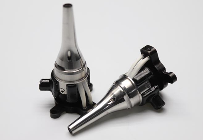
Avantier Ear Endoscope with LED Fiber Optic Illumination. Image Credit: Avantier Inc.
Miniaturizing optical components introduces substantial engineering obstacles. Achieving high optical transmission and resolution with internal lens diameters as small as 2.7 mm, such as those in Avantier ear endoscopes, requires advanced lens design and manufacturing techniques.
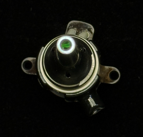
Avantier Ear Endoscope with Ring Fiber Optic Illumination. Image Credit: Avantier Inc.
Clinical applications of lens-based ear endoscopy in otologic diagnosis and treatment
Lens-based ear endoscopes are essential in both diagnosis and treatment. The following is an overview of some of their applications.
1. Diagnostic applications:
- External otitis: High-resolution endoscopy allows in-depth visualization of the entire EAC, aiding in accurately detecting inflammatory changes (erythema, edema), skin lesions, debris, and the presence of cerumen impaction or foreign bodies. In external otitis, the endoscope allows direct observation of canal wall thickening and exudate. For cerumen impaction, the endoscope offers vital insight into the location, size, and consistency of the obstruction, helping doctors identify suitable removal strategies.
- Tympanic membrane assessment: Endoscopic assessment offers a magnified view of the TM, enabling accurate analysis of its integrity, color, contour, and mobility. Subtle indications of middle ear pathology, such as hyperemia, perforation (including location and size), or retraction pockets, are easily visualized. Seeing the tympanic membrane more clearly improves diagnostic precision compared to conventional otoscopy.
- Middle ear diagnostics: In specific cases, especially after tympanic membrane perforation or during surgical examination, rigid endoscopes can be inserted into the middle ear for direct visualization of the ossicular chain, middle ear mucosa, and identification of pathological entities, including ossicular fixation, erosion, or cholesteatoma. Such insights can be invaluable during surgery, helping to shape planning and guide execution.
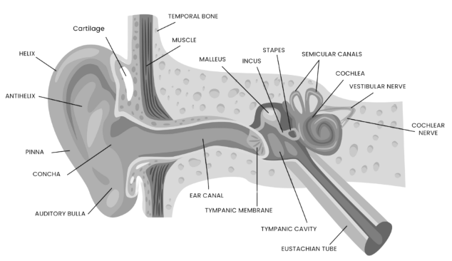
Internal Anatomy of the Human Ear. Image Credit: Avantier Inc.
2. Therapeutic applications:
- Foreign body and cerumen removal: Endoscopic guidance allows for the safe and precise removal of foreign bodies or impacted cerumen from the EAC using specialized micro-instruments introduced alongside the endoscope or via an integrated working channel. Direct visualization reduces the risk of damaging the ear canal and tympanic membrane.
- Endoscopic ear surgery: Lens-based endoscopes are increasingly used as adjuncts or primary visualization instruments in diverse otologic surgeries, such as tympanoplasty, cholesteatoma surgery, and stapedotomy. The endoscope’s enhanced visualization supports accurate tissue manipulation, reduces surgical morbidity, and improves surgical outcomes.
- Postoperative monitoring: Endoscopic examination is essential for postoperative evaluation of the surgical site and can guarantee graft integrity, detect possible complications, and monitor healing.

Clinician Utilizing an Ear Endoscope for Patient Examination. Image Credit: Avantier Inc.
Comparative evaluation with other endoscopic modalities
Otic endoscopes feature design and performance properties that differ from endoscopes used in other anatomical systems (e.g., gastrointestinal, urological). The confined and fragile nature of the ear necessitates a substantially smaller diameter of the endoscope shaft, typically in the millimeter range, to prevent trauma during insertion into the ear canal.
Given the importance of the auditory system, sharp optical resolution and precise illumination are vital for spotting minor changes that could have major effects on hearing.
As a result, otic endoscopes frequently incorporate more advanced optical and illumination systems. The working channels in otic endoscopes are designed for compatibility with micro-instruments required for complex diagnostic and therapeutic maneuvers in the limited surgical space of the ear.
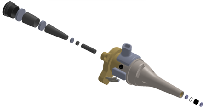
Exploded View of an Ear Endoscope. Image Credit: Avantier Inc.
Future directions and technological developments in ear endoscopy
Significant developments continue to shape the field of ear endoscopy. Rising technologies, such as three-dimensional (3D) imaging modalities and variable magnification, are providing more detailed and spatially precise visualization of otologic structures.
Advances in artificial intelligence are expected to deliver image evaluation algorithms capable of automating the detection and classification of ear pathologies, with the potential to improve diagnostic performance significantly.
Progress in biocompatible materials science is also anticipated to yield endoscope designs that are more flexible and less traumatic, enhancing patient comfort during examinations and procedures. These ongoing innovations are expected further to revolutionize both the diagnosis and treatment of ear diseases.
About Avantier Inc
Avantier is a vertically integrated leader in high-precision custom optics, delivering end-to-end solutions for complex optical challenges. From prototyping to full-scale production, we ensure exceptional quality and repeatability.
Our expertise spans the design and manufacturing of advanced optical components such as aspherical lenses, freeform optics, microscope objective lenses, OAP mirrors, telescopes, SiC optics, optical domes, microlens arrays, large optics, and infrared lenses.
Serving aerospace, biomedical, life sciences, quantum computing, and other advanced technology sectors, Avantier is your trusted one-stop strategic partner for meeting the most stringent optical specifications with precision and reliability.
Sponsored Content Policy: News-Medical.net publishes articles and related content that may be derived from sources where we have existing commercial relationships, provided such content adds value to the core editorial ethos of News-Medical.Net which is to educate and inform site visitors interested in medical research, science, medical devices, and treatments.