The FLUOVIEW FV3000 Series is designed to meet some of the most difficult challenges in modern science. With the high sensitivity and speed required for live cell and tissue imaging, the FV3000 also provides an intuitive and adaptable interface, and is capable of microplate imaging and complex screening protocols.
The series supports complete workflows from live cell 2D-6D (x,y,λ,z,t,p) imaging through image processing, like deconvolution, and analysis. Particular attention has been paid to the needs of cell biology, cancer research, and stem cell research. The FV3000 is optimized for macro to micro imaging of cells, tissues and small organisms.
Features
The FV3000 Series Scan Units
Galvanometer and Galvo/Resonant Hybrid Scanner
Users have their choice of two different types of scan units: galvanometer only with the FV3000 or galvanometer/resonant hybrid with the FV3000RS. The hybrid scan unit has galvanometer scanners for high-precision scanning, as well as a galvo/resonant scanner ideal for high-speed imaging.
Galvanometer scanner enables Evident super resolution technology (FV-OSR) yields resolutions down to 120 nm as well as high signal-to-noise, with precise tornado and multipoint stimulation and 100 ms switching time.
Galvanometer scanning can achieve 16 frames per second at 2X zoom. The resonant scanner is capable of speeds ranging from 30 frames per second at 512 x 512 to 438 frames per second at 512 x 32.
No Compromise between Speed and Field of View
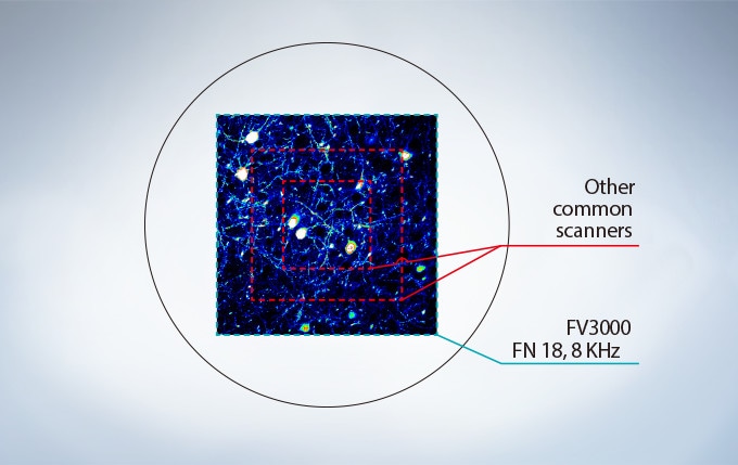
Many high-speed scanning methods restrict the field of view, limiting their usefulness for examining large areas with multiple cells. The FV3000 Series’ resonant scanner maintains a full 1X field of view, even at a video rate of 30 frames per second. Additional speed is generated by clipping the Y axis, even at 438 frames per second.
Most resonant scanners force a trade-off between speed and field of view. FLUOVIEW systems are optimized to maintain the field of view with even signal intensity so dynamic samples (e.g. calcium imaging) can be seen in the broad context of their cells and tissues.
Platelets bound to thrombosis in blood vessel of mouse. Images taken 30 fps in full frame by resonant scanner with 2 CH GaAsP PMTs.
Image data courtesy of Dr. Takuya Hiratsuka, Dr. Michiyuki Matsuda, Graduate School of Biostudies, Kyoto University.
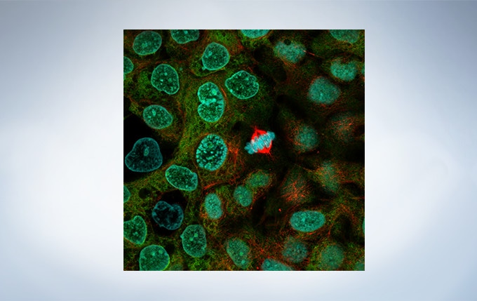
A431 cells fixed with methanol labeled with Abcam Anti-ERK1 + ERK2 antibody (Alexa Fluor 488) ab208564, and Anti-alpha Tubulin antibody (Alexa Fluor 594) ab195889 and DAPI. Sample courtesy of Abcam.
Optimized for Live Cell Imaging
Resonant scanning greatly reduces photobleaching and phototoxicity compared to standard galvanometer scans by preventing the excitation of fluorophores into triplet states that create reactive oxygen species.
These features make live cell experiments more robust and reliable. The FV3000 Series has complete high and low range laser intensity control enabling the system to use the minimum required amount of laser power on samples. The optional Laser Power Monitor provides consistent laser power during long-term time-lapse imaging across multiple days.
Introducing TruSpectral Detection
A Fully Spectral System with Sensitivity and Accuracy
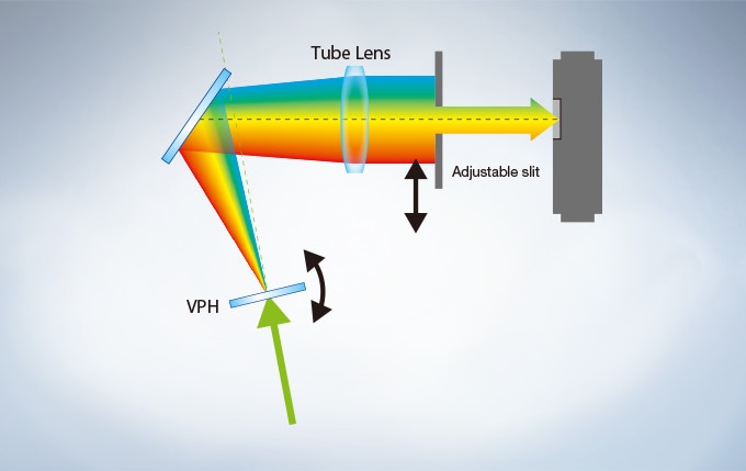
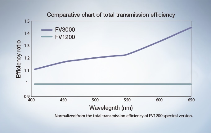
The FV3000 Series employs Evident’ TruSpectral detection concept. Based on patented* Volume Phase Hologram (VPH) transmission and an adjustable slit to control light, the spectral detection in FV3000 and FV3000RS is highly efficient, enabling users to select the detection wavelength of each individual channel to 1 nm.
* US8530824B/JP5541972B/EP2395380A
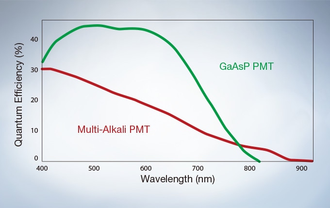
High-Sensitivity Spectral Detector (HSD) with GaAsP Photomultiplier Tubes Enhances Quantum Efficiency
HSD makes it possible to view samples that were too dim to view with conventional equipment. The GaAsP PMT incorporates 2 channels with a maximum quantum efficiency of 45 %, and Peltier cooling reduces background noise by 20 % for high S/N ratio images under exceptionally low excitation light.
Efficient TruSpectral Detection System
The FV3000 Series brings new levels of total system transmission efficiency, enabling every system to be completely spectral, improving overall sensitivity, and improving the signal-to-noise ratio for improved multi-color confocal imaging.
Multichannel TruSpectral Detection with 16-Channel Unmixing
TruSpectral’s efficient design and software enable spectral detectors to run in multichannel mode for both live and postprocessing spectral unmixing with a multichannel lambda mode. Multichannel mode facilitates constant spectral unmixing during live cell experiments, separating complex fluorescence during acquisition. With up to 4 different dynamic ranges from the 4 different channels of array, even bright and dim spectral signals can be separated by adjusting the sensitivity of each detector independently.
Evident Super Resolution (FV-OSR)
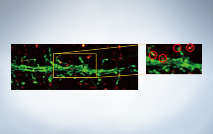
Evident’ widely applicable super resolution method requires no special fluorophores and works for a wide range of samples. Ideal for colocalization analysis, the FV-OSR can acquire 4 fluorescent signals either sequentially or simultaneously with a resolution of approximately 120 nm*, nearly doubling the resolution of typical confocal microscopy.
The system is easy to use with minimal user training and can be added to any confocal system, making the FV-OSR a truly accessible method for achieving super resolution. *Subject to objective magnification, numerical aperture, excitation and emission wavelength, and experiment conditions.
Secondary antibody labels against GFP (Alexa Fluor 488, neurons) and SV2 (Alexa Fluor 565, red). Sample courtesy of Dr. Ed Boyden and Dr. Fei Chen, MIT.

0.5 AU Confocal Image
0.5 AU Confocal Image Deconvolved with cellSens Advanced Deconvolution
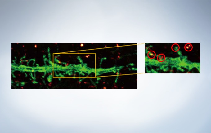
Evident Super Resolution Plus cellSens Advanced Deconvolution. Note clear separation of punctate stains with OSR.