The fully motorized IX83 is the most advanced member of the IX3 series of inverted imaging systems and offers a new level of application flexibility. Choose from the one-deck system with a low, ergonomic stage or the two-deck system with additional expansion capabilities.
Both provide the ability to perform a multitude of imaging applications, from long-term time-lapse imaging and other demanding techniques to routine testing and documentation. Users can mold the components and controls to best suit their workflow.
Features
Expandable to Meet Growing Research Needs
The fully-motorized IX83 is designed to satisfy a variety of research needs. With additional modules providing expanded functionality, both microscope options enable a multitude of imaging techniques, ranging from casual documentation to long-term time-lapse imaging and other demanding techniques.
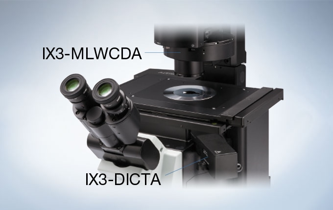
The unique open frame of the IX83 provides ready access to the light path, thus making it easy to add or change modules. A variety of deck modules can be easily exchanged to add or remove functions as needed. With a simple slide-in design, the IX3-ZDC2 Z drift compensator module can be easily added to any IX83 system to maintain continuous focus throughout an extended time-lapse experiment.
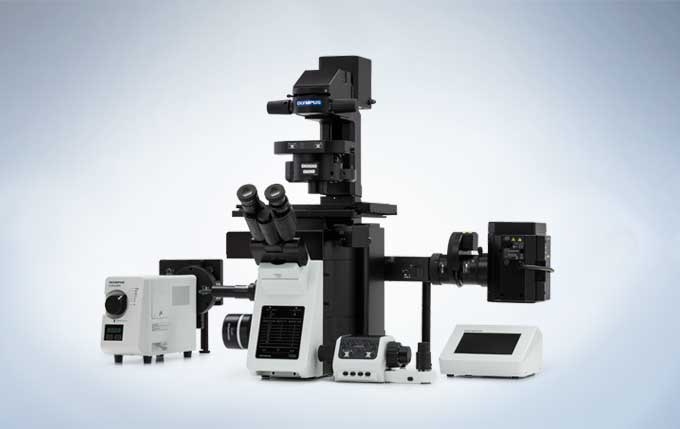
IX83: Two-deck System
Enables high-speed, fully automated device selection during live cell research and advanced image acquisition. The two-deck configuration provides excellent expandability.
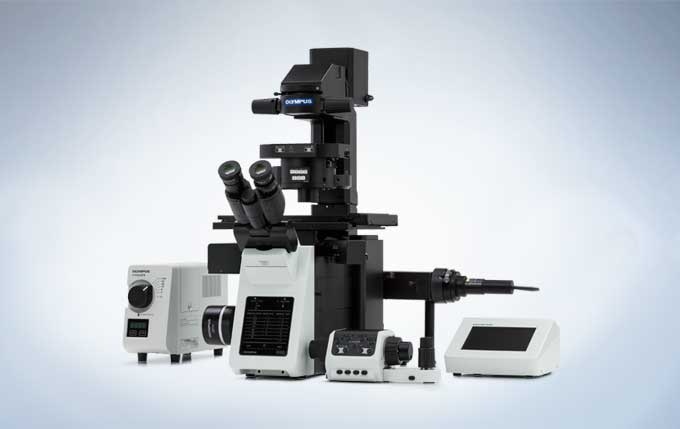
IX83: One-deck System
An intelligent, motorized microscope featuring a large field number (FN22, left side port) and IX3-ZDC2 compatibility, thus creating a new standard for live cell imaging.
Reliable, Clear and Bright High-Resolution Images
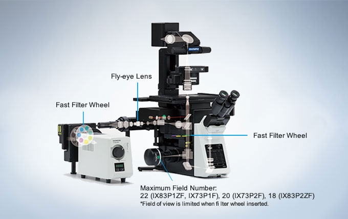
Evident UIS2 infinity-corrected optics ensure high optical transmittance with a broad range of objectives. UIS2 optics feature wide chromatic correction and enable high resolution, high S/N primary images regardless of the observation method. The wide field of view and Fly-Eye lens system provide uniform fluorescence images and enable the use of sCMOS cameras with large sensors.
Excellent Image Quality
Apochromatic Objectives Enable High-Resolution Phase Contrast and Fluorescence Observation
Phase contrast apochromatic objectives (UPLSAPO100XOPH, PLAPON60XOPH) enable high-precision imaging free from image shift, even during simultaneous phase contrast and fluorescence observation. This eliminates the need to change objectives when switching observation methods.
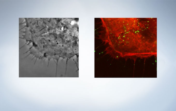
HeLa cell expressing mCherry-actin (Image data courtesy of: Tomonobu Watanabe, Ph.D. Laboratory for Comprehensive Bioimaging, RIKEN Quantitative Biology Center)
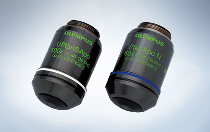
UPLSAPO100×OPH, PLAPON60×OPH
Silicone Objectives* Enable High-Resolution Observation Deep into Live Cells
Evident offers three high-NA silicone immersion objectives:UPLSAPO30XS, UPLSAPO40XS, and UPLSAPO60XS. The refractive index of silicone oil (Refractive index: ne≈1.40) is close to that of living tissue (Refractive index: ne≈1.38), enabling high-resolution observation deep inside living tissue with minimal spherical aberration caused by refractive index mismatch. Silicone oil does not dry out or harden, so there is never a need to readminister the oil, making it ideal for extended time-lapse observations.
*Use dedicated silicone oil.
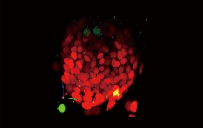
3D reconstruction images of a live sphere made of NMuMG/Fucci2 cells. Confocal images were acquired by using an Evident FV1000 confocal microscope. (Red: cell cycle G1 phase, Green: cell cycle S/G2/M phase) Image data courtesy of: Asako Sakaue-Sawano, Ph.D. Atsushi Miyawaki, M.D., Ph.D. Laboratory for Cell Function Dynamics, Advanced Technology Development Core, RIKEN Brain Science Institute
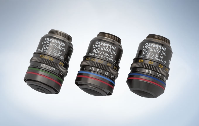
UPLSAPO30XS, UPLSAPO40XS and UPLSAPO60XS
Special Objective Available for iPS/ES and Floating Cell Observation
This high-NA phase contrast objective (UCPLFLN20XPH) is especially suited for observation using plastic dishes. It enables high-resolution observation of the cell proliferation process and delivers improved contrast across a wide area.
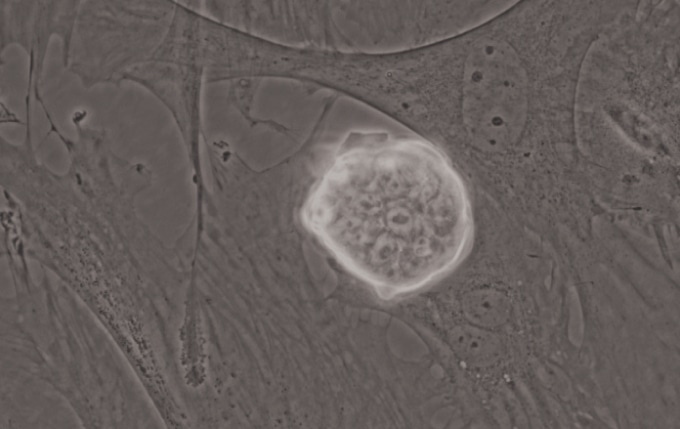
iPS-cell expressing Nanog reporter (GFP) Image data courtesy of: Tomonobu Watanabe, Ph.D. Laboratory for Comprehensive Bioimaging, RIKEN Quantitative Biology Center
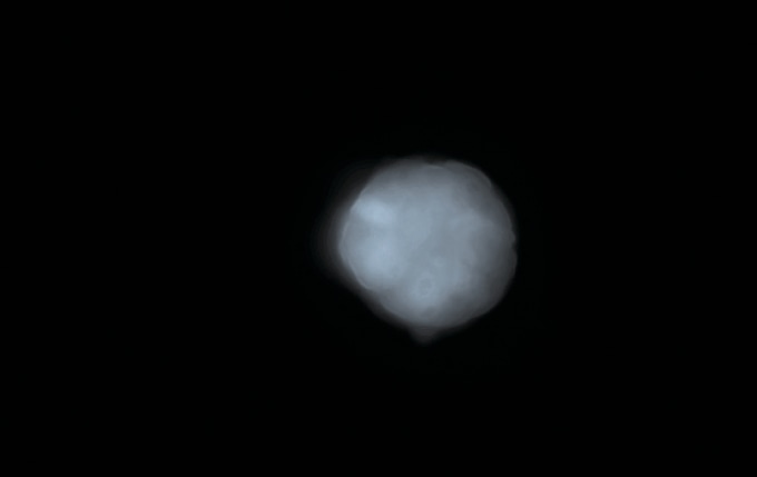
iPS-cell expressing Nanog reporter (GFP) Image data courtesy of: Tomonobu Watanabe, Ph.D. Laboratory for Comprehensive Bioimaging, RIKEN Quantitative Biology Center
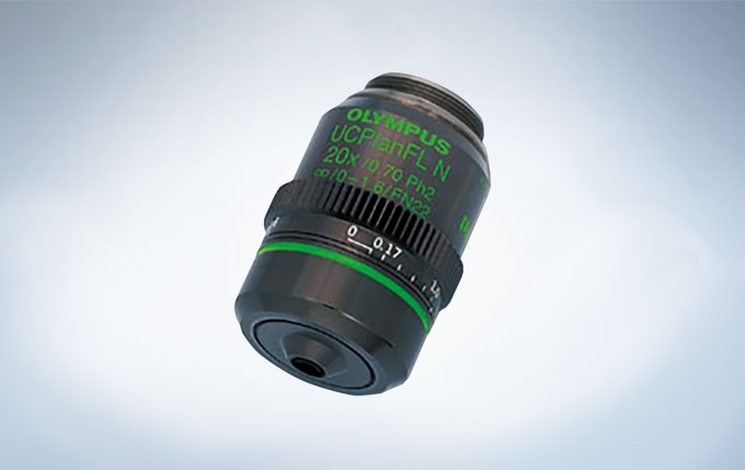
UCPLFLN20×PH
Bright, Uniform Fluorescence Illumination
The fluorescence illuminator (IX3-RFALFE) incorporates a Fly-Eye lens system to provide even light distribution. This provides bright and even illumination to the entire field, including the periphery of the visual field.
With Fly-Eye Lens System
Without Fly-Eye Lens System
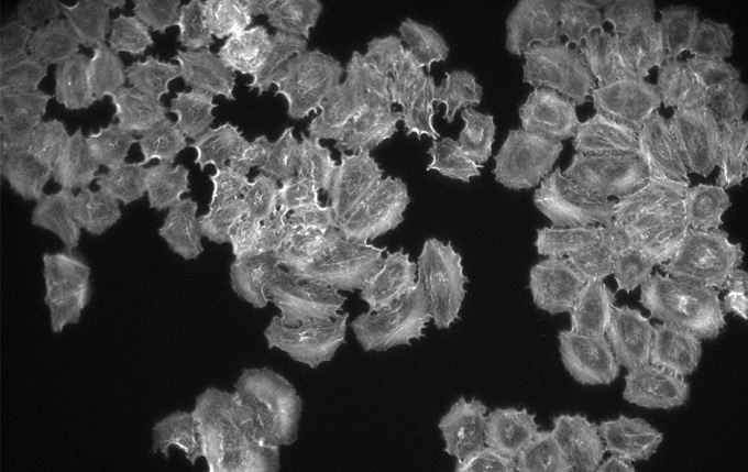
High S/N Fluorescence Mirror Units for Efficient Signal Detection
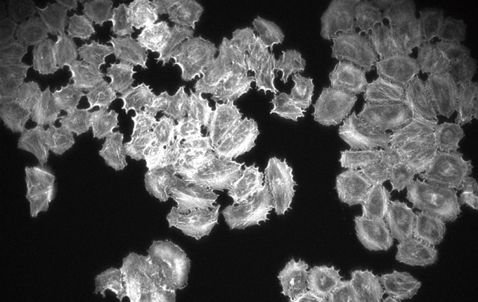
All fluorescence mirror units feature filters treated with a specially-developed coating that minimizes noise by absorbing more than 99% of stray light. The outstanding performance and high transmittance of the mirror units ensure efficient fluorescence signal detection.
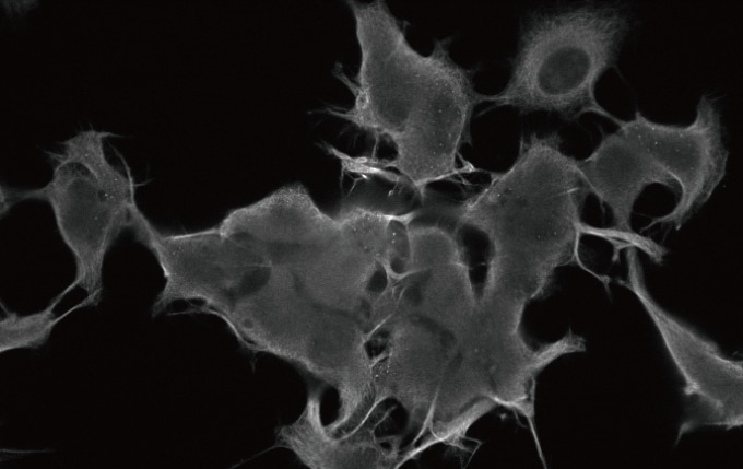
Image Captured with a Conventional FL Mirror Unit

Images Captured with New FL Mirror Units
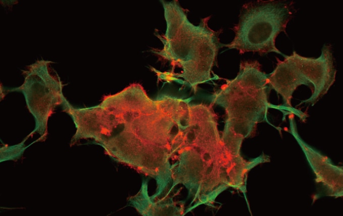
Images Captured with New FL Mirror Units
Ideally Suited for Live Cell Imaging
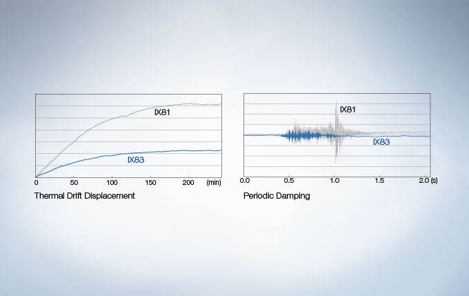
Easily capture dynamic cellular processes with the IX83, an imaging system designed for remarkable time-lapse imaging. With new frame architecture and focus drive design, the IX3 system offers enhanced rigidity that reduces the impact of vibration and temperature.
It maintains desired positions along the X, Y, and Z axes to facilitate reliable time-lapse and multipoint imaging.When combined with the Evident IX3-SSU ultrasonic stage and Z drift compensator (IX3-ZDC2) , the IX83 is perfectly suited for capturing high-precision, multipoint time-lapse images that are never out of focus or misaligned. Box-type and stage-top incubators are also available to maintain the viability of live cells during time-lapse experimentation.
Easily Capture Time-Lapse Images
Easily Capture Time-Lapse Images
Easily Capture Time-Lapse Images