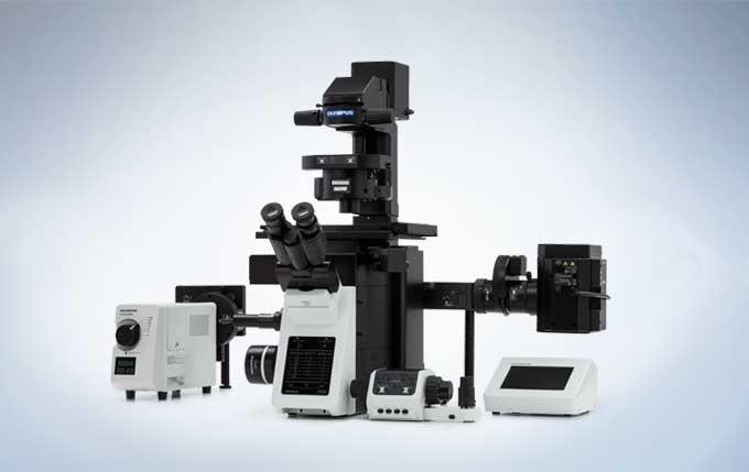
The IX73 inverted microscope system sets new standards in advanced live cell imaging with its compact frame, outstanding optical performance and exceptional flexibility. Manual encoded or semi-motorized options enable a variety of component combinations.
The IX73 is available as a one-deck system with a low, ergonomic stage or as a two-deck system with additional expansion capabilities. Both provide the ability to perform a multitude of imaging applications, from fast advanced fluorescence imaging and other demanding techniques to routine testing and documentation.
Features
Expandable to Meet Growing Research Needs
The semi-motorized IX73 is designed to satisfy a variety of research needs. With two deck options and additional modules for expanded functionality, the IX73 is perfectly suited for a changing research environment.
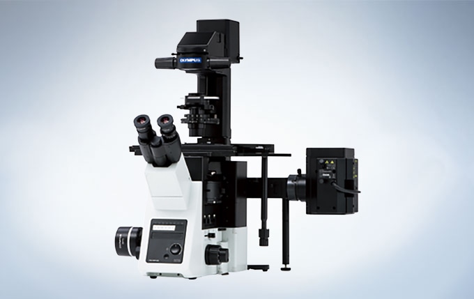
IX73: Two-deck System
The modular IX73 two-deck system can be combined with coded or motorized units for maximum expandability.
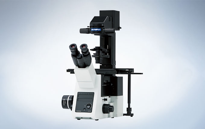
IX73: One-deck System
A microscope designed with an emphasis on efficiency. Ideal for documentation, routine testing and other tasks.
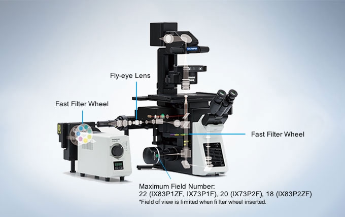
Reliable, Clear and Bright High-Resolution Images
Evident UIS2 infinity-corrected optics ensure high optical transmittance with a broad range of objectives. UIS2 optics feature wide chromatic correction and enable high resolution, high S/N primary images regardless of the observation method. The wide field of view and Fly-Eye lens system provide uniform fluorescence images and enable the use of sCMOS cameras with large sensors.
Excellent Image Quality
Apochromatic Objectives Enable High-Resolution Phase Contrast and Fluorescence Observation
Phase contrast apochromatic objectives (UPLSAPO100XOPH, PLAPON60XOPH) enable high-precision imaging free from image shift, even during simultaneous phase contrast and fluorescence observation. This eliminates the need to change objectives when switching observation methods.
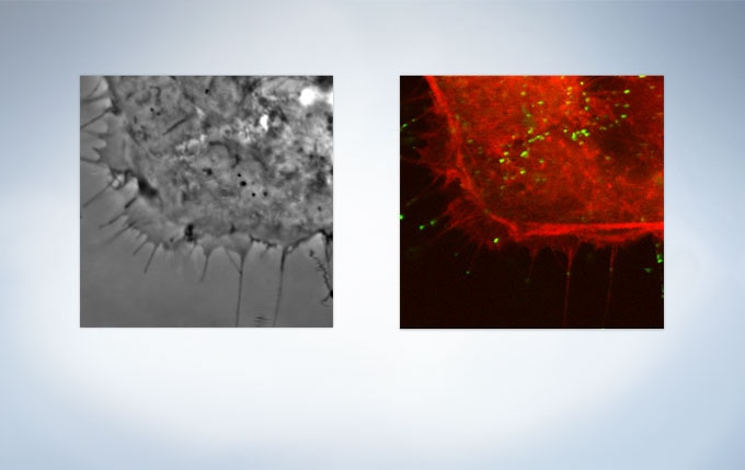
HeLa cell expressing mCherry-actin (Image data courtesy of: Tomonobu Watanabe, Ph.D. Laboratory for Comprehensive Bioimaging, RIKEN Quantitative Biology Center)
IX83:Two-deck System
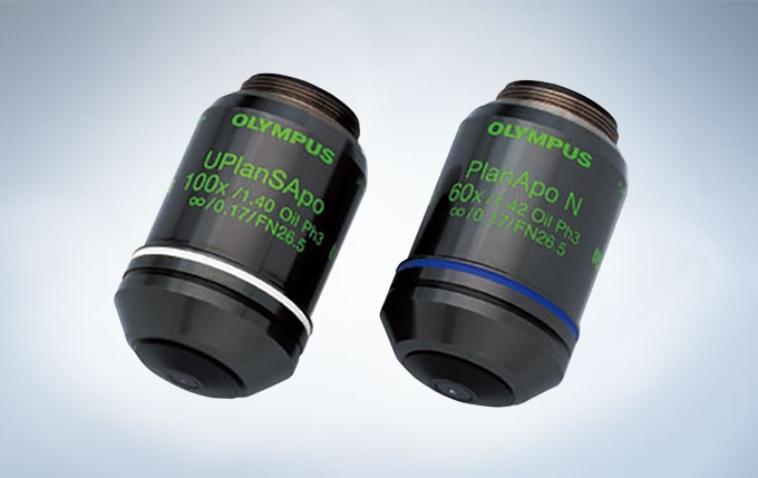
UPLSAPO100×OPH, PLAPON60×OPH
Silicone Objectives* Enable High-Resolution Observation Deep into Live Cells
Evident offers three high-NA silicone immersion objectives:UPLSAPO30XS, UPLSAPO40XS, and UPLSAPO60XS. The refractive index of silicone oil (Refractive index: ne≈1.40) is close to that of living tissue (Refractive index: ne≈1.38), enabling high-resolution observation deep inside living tissue with minimal spherical aberration caused by refractive index mismatch. Silicone oil does not dry out or harden, so there is never a need to readminister the oil, making it ideal for extended time-lapse observations.
*Use dedicated silicone oil.
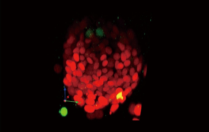
3D reconstruction images of a live sphere made of NMuMG/Fucci2 cells. Confocal images were acquired by using an Evident FV1000 confocal microscope. (Red: cell cycle G1 phase, Green: cell cycle S/G2/M phase) Image data courtesy of: Asako Sakaue-Sawano, Ph.D. Atsushi Miyawaki, M.D., Ph.D. Laboratory for Cell Function Dynamics, Advanced Technology Development Core, RIKEN Brain Science Institute
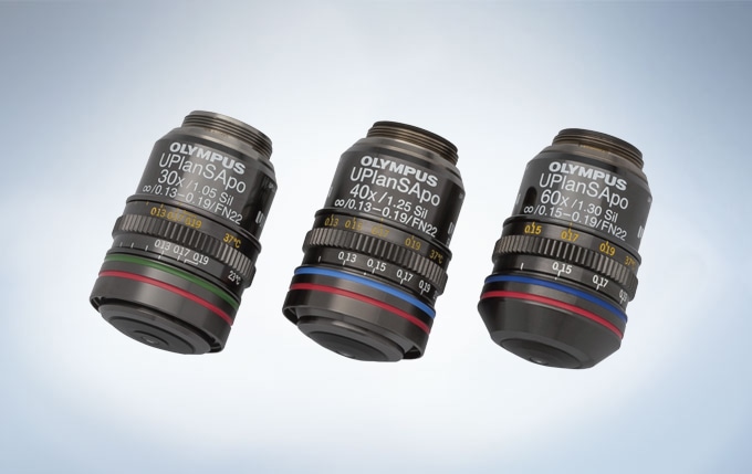
UPLSAPO30XS, UPLSAPO40XS and UPLSAPO60XS
Special Objective Available for iPS/ES and Floating Cell Observation
This high-NA phase contrast objective (UCPLFLN20XPH) is especially suited for observation using plastic dishes. It enables high-resolution observation of the cell proliferation process and delivers improved contrast across a wide area.
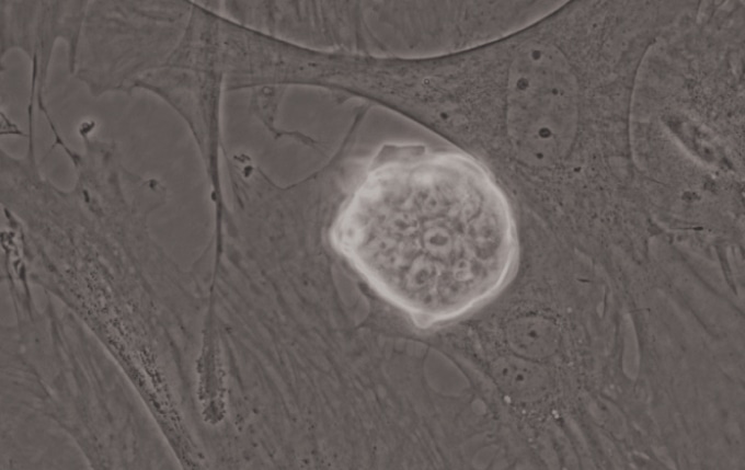
iPS-cell expressing Nanog reporter (GFP) Image data courtesy of: Tomonobu Watanabe, Ph.D. Laboratory for Comprehensive Bioimaging, RIKEN Quantitative Biology Center
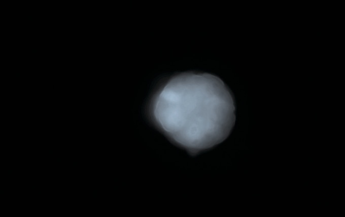
iPS-cell expressing Nanog reporter (GFP) Image data courtesy of: Tomonobu Watanabe, Ph.D. Laboratory for Comprehensive Bioimaging, RIKEN Quantitative Biology Center
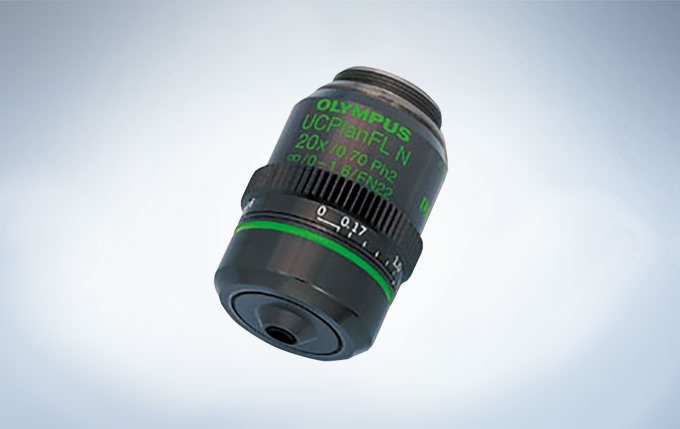
UCPLFLN20×PH